Copyright
©The Author(s) 2018.
World J Clin Cases. Nov 26, 2018; 6(14): 830-835
Published online Nov 26, 2018. doi: 10.12998/wjcc.v6.i14.830
Published online Nov 26, 2018. doi: 10.12998/wjcc.v6.i14.830
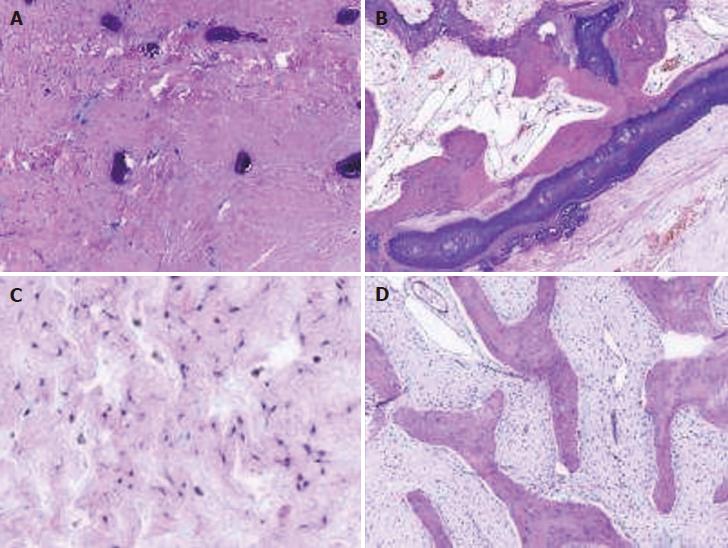
Figure 3 Pathological reports.
A: Calcified woven bone can be seen (HE; 40 ×); B: No osteoblast and osteoclast hyperplasia can be seen (HE; 40 ×); C: It shows Fibroblast and Mucoid matrix (HE; 40 ×); D: It shows fibro-osseous lesions, with discontinuity of the trabecular bone and calcification (HE; 40 ×). HE: Hematoxylin-eosin.
- Citation: Liu YB, Zou TM. Giant monostotic osteofibrous dysplasia of the ilium: A case report and review of literature. World J Clin Cases 2018; 6(14): 830-835
- URL: https://www.wjgnet.com/2307-8960/full/v6/i14/830.htm
- DOI: https://dx.doi.org/10.12998/wjcc.v6.i14.830


