Copyright
©The Author(s) 2017.
World J Clin Cases. Feb 16, 2017; 5(2): 35-39
Published online Feb 16, 2017. doi: 10.12998/wjcc.v5.i2.35
Published online Feb 16, 2017. doi: 10.12998/wjcc.v5.i2.35
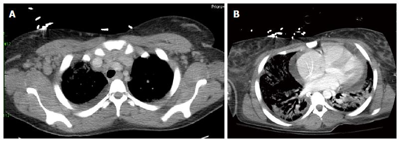
Figure 1 Computerized tomography chest.
A: Computerized tomography (CT) chest demonstrating significant axillary lymphadenopathy; B: CT chest revealing bilateral lung consolidation, and a moderate pericardial effusion.
- Citation: Barbat B, Jhaj R, Khurram D. Fatality in Kikuchi-Fujimoto disease: A rare phenomenon. World J Clin Cases 2017; 5(2): 35-39
- URL: https://www.wjgnet.com/2307-8960/full/v5/i2/35.htm
- DOI: https://dx.doi.org/10.12998/wjcc.v5.i2.35


