Copyright
©The Author(s) 2015.
World J Clin Cases. Jun 16, 2015; 3(6): 533-537
Published online Jun 16, 2015. doi: 10.12998/wjcc.v3.i6.533
Published online Jun 16, 2015. doi: 10.12998/wjcc.v3.i6.533
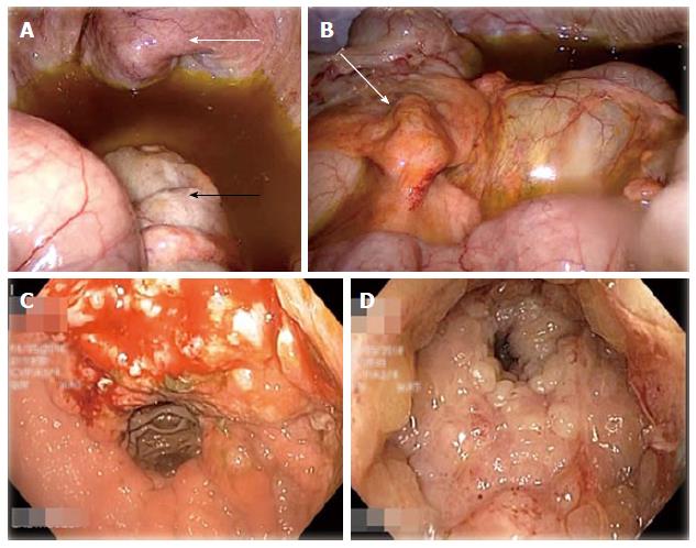
Figure 1 Laparoscopy examination.
A: Laparoscopic view of the pelvic organs; Bladder (white arrow) and rectum (black arrow) look grossly normal. Moderate amount of greenish ascites seen; B: Transverse colon serosa normal looking except for thickened appendices epiploicae (white arrow); C: Gastric tumour seen at lesser curve on EGD; D: Extensive circumferential rectal mucosal tumour with luminal narrowing.
- Citation: Seow-En I, Seow-Choen F. Intestinal type gastric adenocarcinoma with unusual synchronous metastases to the colorectum and bladder. World J Clin Cases 2015; 3(6): 533-537
- URL: https://www.wjgnet.com/2307-8960/full/v3/i6/533.htm
- DOI: https://dx.doi.org/10.12998/wjcc.v3.i6.533


