Copyright
©The Author(s) 2015.
World J Clin Cases. Dec 16, 2015; 3(12): 1011-1016
Published online Dec 16, 2015. doi: 10.12998/wjcc.v3.i12.1011
Published online Dec 16, 2015. doi: 10.12998/wjcc.v3.i12.1011
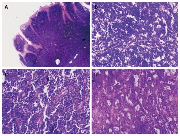
Figure 3 Microscopically the morphological picture.
A: Monotonous population of basophilic round cells with minimal stroma beneath the surface epithelium (4 ×); B: Homogenous tumor cells with basophilic nuclei and minimal cytoplasm separated by thin fibrous septa (10 ×); C: Tumor cells showing coarse chromatin and multiple nucleoli indicating high mitotic index (10 ×); D: Macrophages with foamy cytoplasm are seen scattered amongst tumor cells (10 ×).
- Citation: Patankar S, Venkatraman P, Sridharan G, Kane S. Burkitt’s lymphoma of maxillary gingiva: A case report. World J Clin Cases 2015; 3(12): 1011-1016
- URL: https://www.wjgnet.com/2307-8960/full/v3/i12/1011.htm
- DOI: https://dx.doi.org/10.12998/wjcc.v3.i12.1011


