
















©2014 Baishideng Publishing Group Inc.
World J Clin Cases. May 16, 2014; 2(5): 151-156
Published online May 16, 2014. doi: 10.12998/wjcc.v2.i5.151
Published online May 16, 2014. doi: 10.12998/wjcc.v2.i5.151
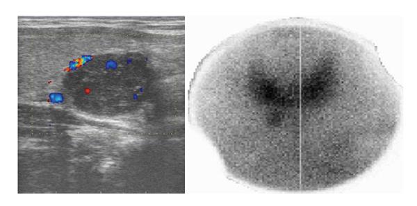
Figure 1 Doppler ultrasound of parathyroid gland showing well-defined hypoechoic mass with color signals of few surrounding vascular structures and Tc-99m sestamibi planar scan with pinhole collimator, 10 min post injection.
- Citation: Baretić M, Tomić Brzac H, Dobrenić M, Jakovčević A. Parathyroid carcinoma in pregnancy. World J Clin Cases 2014; 2(5): 151-156
- URL: https://www.wjgnet.com/2307-8960/full/v2/i5/151.htm
- DOI: https://dx.doi.org/10.12998/wjcc.v2.i5.151