Copyright
©2014 Baishideng Publishing Group Inc.
World J Clin Cases. May 16, 2014; 2(5): 142-145
Published online May 16, 2014. doi: 10.12998/wjcc.v2.i5.142
Published online May 16, 2014. doi: 10.12998/wjcc.v2.i5.142
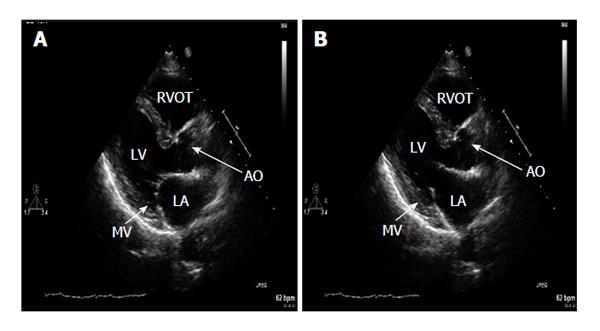
Figure 1 Transthoracic 2D parasternal long axis.
A: Systole shows diffuse echo dense signal significantly confined to the walls of the left atrium indicated by arrow. No mitral valve (MV) or mitral annulus involvement. Valve closes normally during systole; B: Diastole shows adequate MV opening. LA: Left atrium; AO: Aortic root.
- Citation: Jones C, Lodhi AM, Cao LB, Chagarlamudi AK, Movahed A. Atrium of stone: A case of confined left atrial calcification without hemodynamic compromise. World J Clin Cases 2014; 2(5): 142-145
- URL: https://www.wjgnet.com/2307-8960/full/v2/i5/142.htm
- DOI: https://dx.doi.org/10.12998/wjcc.v2.i5.142


