Copyright
©2014 Baishideng Publishing Group Co.
World J Clin Cases. Mar 16, 2014; 2(3): 67-71
Published online Mar 16, 2014. doi: 10.12998/wjcc.v2.i3.67
Published online Mar 16, 2014. doi: 10.12998/wjcc.v2.i3.67
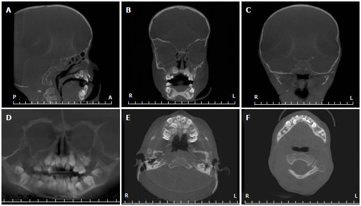
Figure 3 Craniofacial features of the Hutchinson-Gilford progeria syndrome patient detected by cone beam computed tomography.
A, B: On sagittal (A) and coronal (B) images, hypoplasia of the sphenoid, frontal and maxillary sinuses was evident; C: This view depicts the temporomandibular joint alteration, which was characterized by flattening of the condyle and glenoid fossa and bilateral hypoplasia of the condyles; D: Panoramic view of the cone beam computed tomography (CBCT) revealing impaction of several permanent molars and hypodontia of the premolars; E, F: Axial slices of the CBCT showed malocclusion and lingual eruption of permanent teeth.
- Citation: Alves DB, Silva JM, Menezes TO, Cavaleiro RS, Tuji FM, Lopes MA, Zaia AA, Coletta RD. Clinical and radiographic features of Hutchinson-Gilford progeria syndrome: A case report. World J Clin Cases 2014; 2(3): 67-71
- URL: https://www.wjgnet.com/2307-8960/full/v2/i3/67.htm
- DOI: https://dx.doi.org/10.12998/wjcc.v2.i3.67


