
















©2014 Baishideng Publishing Group Inc.
World J Clin Cases. Dec 16, 2014; 2(12): 907-911
Published online Dec 16, 2014. doi: 10.12998/wjcc.v2.i12.907
Published online Dec 16, 2014. doi: 10.12998/wjcc.v2.i12.907
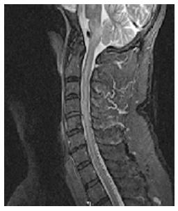
Figure 1 Magnetic resonance imaging of the cervical spine showing increased T2/STIR signal intensity in the pons, medulla, and upper cervical spine and multiple small flow voids in the dorsal cervicothoracic spine suggestive of dural arteriovenous malformation.
- Citation: Gross R, Ali R, Kole M, Dorbeistein C, Jayaraman MV, Khan M. Tentorial dural arteriovenous fistula presenting as myelopathy: Case series and review of literature. World J Clin Cases 2014; 2(12): 907-911
- URL: https://www.wjgnet.com/2307-8960/full/v2/i12/907.htm
- DOI: https://dx.doi.org/10.12998/wjcc.v2.i12.907