Copyright
©2014 Baishideng Publishing Group Inc.
World J Clin Cases. Nov 16, 2014; 2(11): 623-641
Published online Nov 16, 2014. doi: 10.12998/wjcc.v2.i11.623
Published online Nov 16, 2014. doi: 10.12998/wjcc.v2.i11.623
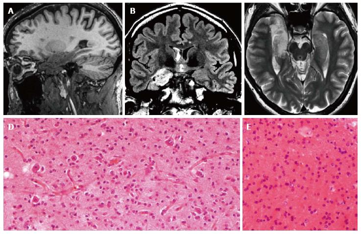
Figure 12 Gangliogliomas and focal cortical dysplasia IIa.
Sagittal 3D T1-w (A), coronal FLAIR T2-w (B) and axial T2-w (C) reveal an inhomogeneous mass, involving the right hippocampus and the temporal pole. Due to the size of the tumor, the associated dysplasia is not clearly visible. Histological examination demonstrates the presence of a glioneuronal tumor with small ganglion cells in a desmoplastic stroma (D) and of dysmorphic neurons in the adjacent cortex (focal cortical dysplasia Type IIa) (E).
- Citation: Giulioni M, Marucci G, Martinoni M, Marliani AF, Toni F, Bartiromo F, Volpi L, Riguzzi P, Bisulli F, Naldi I, Michelucci R, Baruzzi A, Tinuper P, Rubboli G. Epilepsy associated tumors: Review article. World J Clin Cases 2014; 2(11): 623-641
- URL: https://www.wjgnet.com/2307-8960/full/v2/i11/623.htm
- DOI: https://dx.doi.org/10.12998/wjcc.v2.i11.623


