
















©2014 Baishideng Publishing Group Inc.
World J Clin Cases. Oct 16, 2014; 2(10): 581-586
Published online Oct 16, 2014. doi: 10.12998/wjcc.v2.i10.581
Published online Oct 16, 2014. doi: 10.12998/wjcc.v2.i10.581
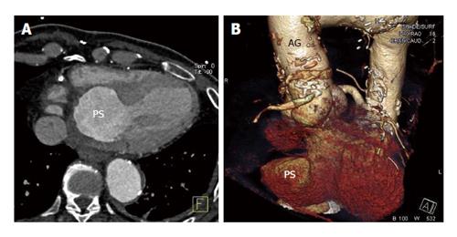
Figure 2 Cardiac computed tomographic angiography (A) and volume rendering (B): Left ventricle posterior wall pseudoaneurysm.
PS: Pseudoaneurysm; AG: Aortic graft.
- Citation: Petrou E, Vartela V, Kostopoulou A, Georgiadou P, Mastorakou I, Kogerakis N, Sfyrakis P, Athanassopoulos G, Karatasakis G. Left ventricular pseudoaneurysm formation: Two cases and review of the literature. World J Clin Cases 2014; 2(10): 581-586
- URL: https://www.wjgnet.com/2307-8960/full/v2/i10/581.htm
- DOI: https://dx.doi.org/10.12998/wjcc.v2.i10.581