Copyright
©2014 Baishideng Publishing Group Co.
World J Clin Cases. Jan 16, 2014; 2(1): 20-23
Published online Jan 16, 2014. doi: 10.12998/wjcc.v2.i1.20
Published online Jan 16, 2014. doi: 10.12998/wjcc.v2.i1.20
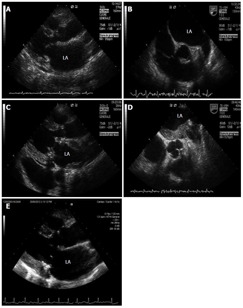
Figure 1 Echocardiographic image.
A: Left parasternal long axis transthoracic echocardiography; B: Upper transesophageal views of the aortic valve in short axis; C: Left parasternal long axis transthoracic echocardiography; D: Upper transesophageal views of the aortic valve in short axis; E: Left parasternal long axis transthoracic echocardiography. LA: left atrium.
- Citation: Rosa GM, Parodi A, Dorighi U, Carbone F, Mach F, Quercioli A, Montecucco F, Vuilleumier N, Balbi M, Brunelli C. Left atrial thrombosis in an anticoagulated patient after bioprosthetic valve replacement: Report of a case. World J Clin Cases 2014; 2(1): 20-23
- URL: https://www.wjgnet.com/2307-8960/full/v2/i1/20.htm
- DOI: https://dx.doi.org/10.12998/wjcc.v2.i1.20


