
















©The Author(s) 2025.
World J Clin Cases. Oct 16, 2025; 13(29): 108046
Published online Oct 16, 2025. doi: 10.12998/wjcc.v13.i29.108046
Published online Oct 16, 2025. doi: 10.12998/wjcc.v13.i29.108046
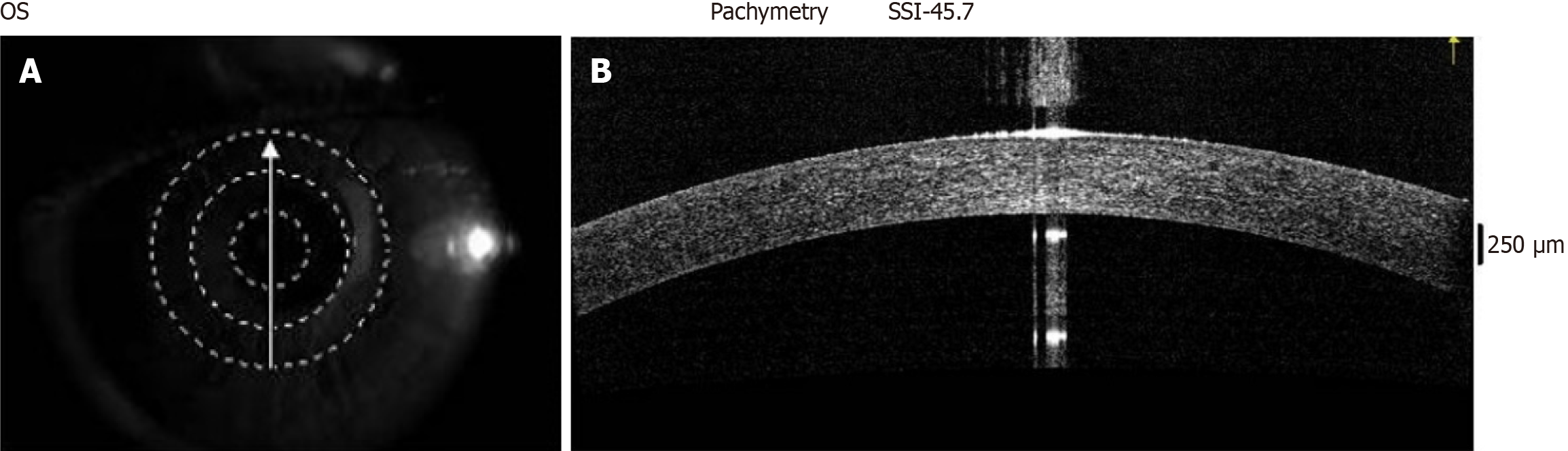
Figure 2 Anterior segment optical coherence tomography image depicting the air-tear film interface, hyporeflective cornea epithelium, and hyperreflective intracorneal tissue.
A: Optical coherence tomography interface as seen on monitor showing direction of scan; B: Single tomogram of the cornea and anterior chamber. OS: Outer segment; SSI: Signal strength index.
- Citation: Enaholo E, Okoye G, Musa M, Suleman A, Ojo O, Foti R, D’Esposito F, Giglio R, Tognetto D, Gagliano C, Zeppieri M. High-resolution optical coherence tomography for screening ocular surface tumors: Historical markers and future directions. World J Clin Cases 2025; 13(29): 108046
- URL: https://www.wjgnet.com/2307-8960/full/v13/i29/108046.htm
- DOI: https://dx.doi.org/10.12998/wjcc.v13.i29.108046