Copyright
©The Author(s) 2025.
World J Clin Cases. Sep 26, 2025; 13(27): 108923
Published online Sep 26, 2025. doi: 10.12998/wjcc.v13.i27.108923
Published online Sep 26, 2025. doi: 10.12998/wjcc.v13.i27.108923
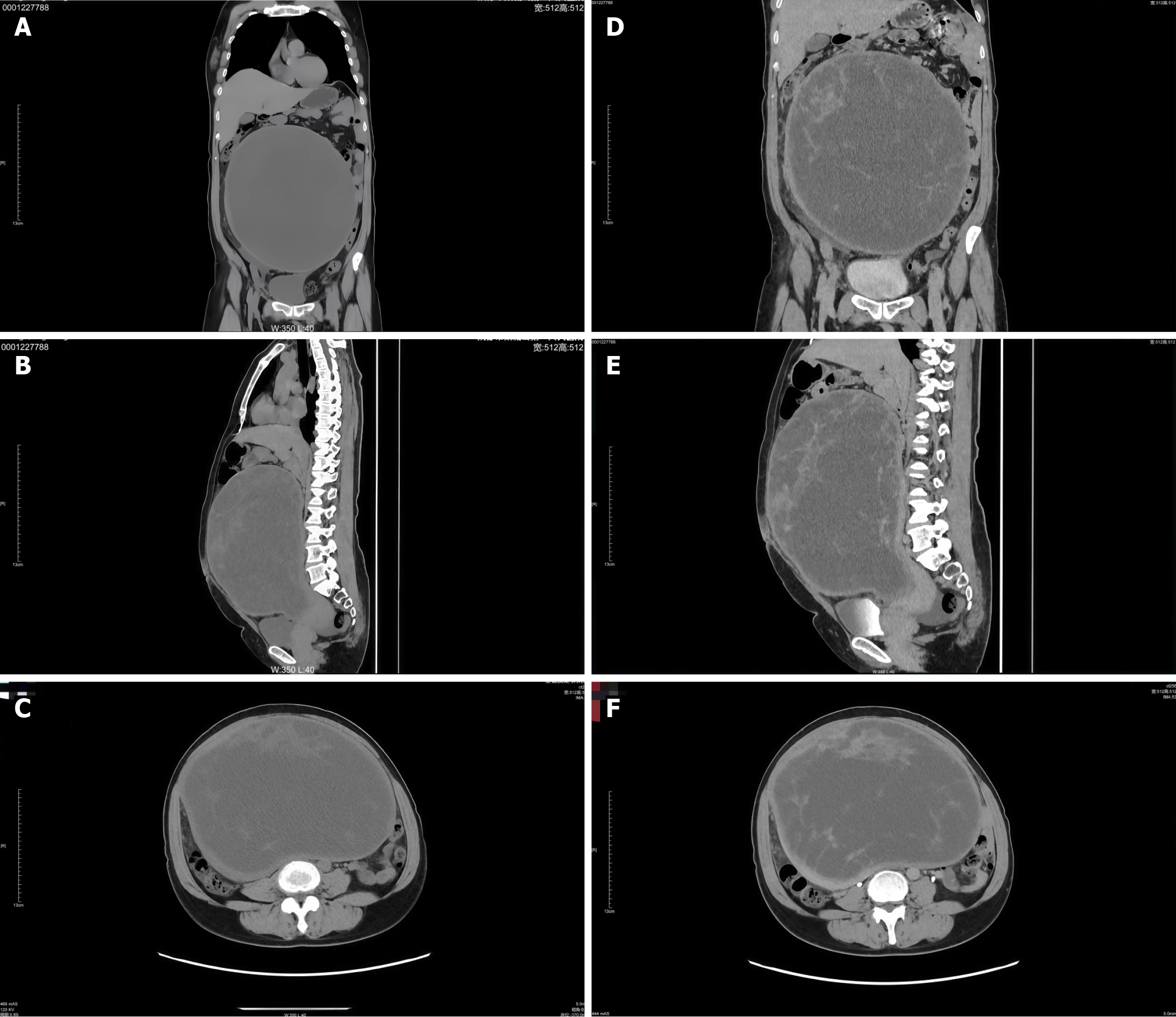
Figure 1 Computed tomography images of the pelvic cavity mass.
A: Coronal section without contrast; B: Sagittal section without contrast; C: Axial section without contrast; D: Coronal section with contrast enhancement; E: Sagittal section with contrast enhancement; F: Axial section with contrast enhancement.
- Citation: Huang X, Yao Q, Wu YC, Liu SC. Diagnostic and surgical management of giant broad ligament myoma with cystic degeneration: A case report. World J Clin Cases 2025; 13(27): 108923
- URL: https://www.wjgnet.com/2307-8960/full/v13/i27/108923.htm
- DOI: https://dx.doi.org/10.12998/wjcc.v13.i27.108923


