Copyright
©The Author(s) 2025.
World J Clin Cases. Aug 26, 2025; 13(24): 105244
Published online Aug 26, 2025. doi: 10.12998/wjcc.v13.i24.105244
Published online Aug 26, 2025. doi: 10.12998/wjcc.v13.i24.105244
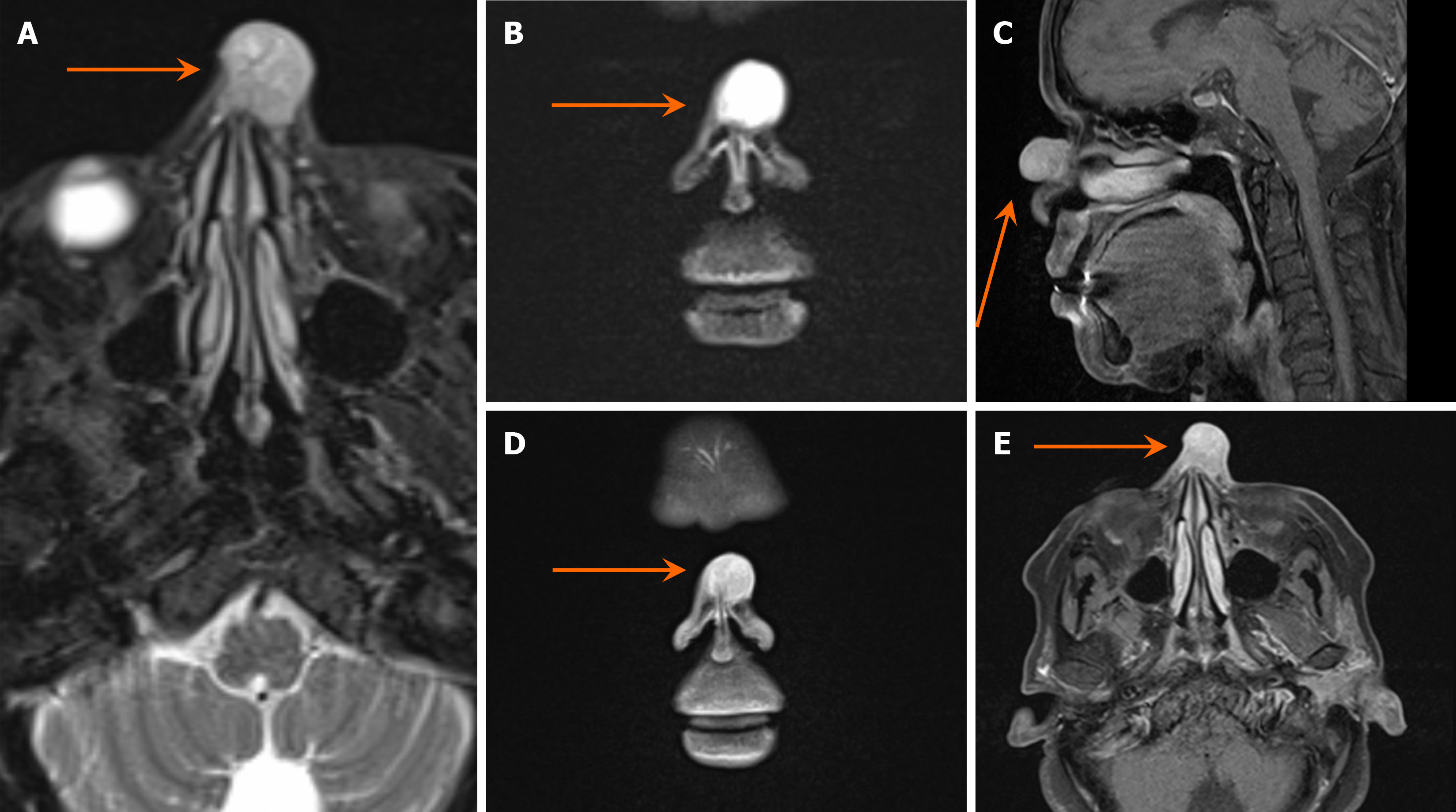
Figure 3 Multiple sequential multiplanar magnetic resonance images of the paranasal sinuses, with IV contrast showing a midline cutaneous pedunculated nasal mass that measured 1.
8 cm × 1.9 cm × 1.9 cm in the maximum axial and cell carcinoma dimensions, respectively. A-B: Heterogenous, predominantly high in T2; C-E: Isointense T1 signal with homogenous contrast enhancement in a post-contrast study.
- Citation: Abuharb AIA, Alzamil AF, Alqarni KS, Alsudays AM, Alqahtani SM, Alahmadi RM, Mutairy ASA, Alghamdi FR. Merkel cell carcinoma presenting as a nasal dorsum mass: A case report and literature review. World J Clin Cases 2025; 13(24): 105244
- URL: https://www.wjgnet.com/2307-8960/full/v13/i24/105244.htm
- DOI: https://dx.doi.org/10.12998/wjcc.v13.i24.105244


