Copyright
©The Author(s) 2025.
World J Clin Cases. Aug 6, 2025; 13(22): 104924
Published online Aug 6, 2025. doi: 10.12998/wjcc.v13.i22.104924
Published online Aug 6, 2025. doi: 10.12998/wjcc.v13.i22.104924
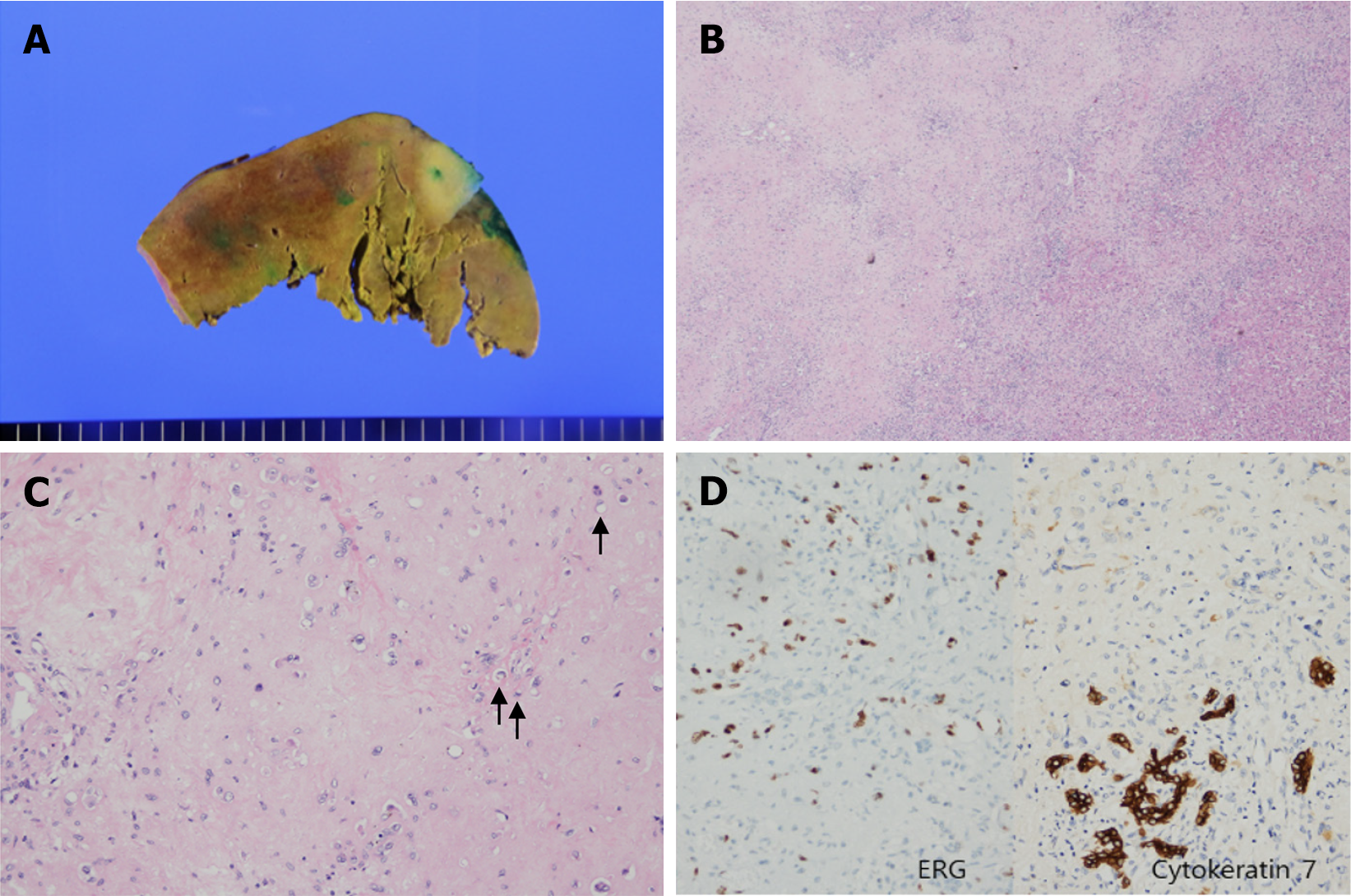
Figure 3 Pathologic findings of hepatic epithelioid hemangioendothelioma.
A: The cut surface shows an ill-defined whitish mass; B: The tumor demonstrates hypercellularity with margins indistinct from the surrounding tissue (H&E stain, × 20); C: Tumor cells are arranged individually or in cords within a myxohyaline stroma, with frequent intracytoplasmic vacuoles (black arrows) (H&E stain, ×200); D: Immunohistochemistry reveals strong reactivity for ERG, indicating a vascular endothelial origin, and also shows immunoreactivity for Cytokeratin 7. The CK7-positive staining pattern appeared morphologically and resembled the intrahepatic bile ducts.
- Citation: Shin SH, Koh YS, Song S. Hepatic epithelioid hemangioendothelioma managed with minimally invasive surgery: A case report. World J Clin Cases 2025; 13(22): 104924
- URL: https://www.wjgnet.com/2307-8960/full/v13/i22/104924.htm
- DOI: https://dx.doi.org/10.12998/wjcc.v13.i22.104924


