Copyright
©The Author(s) 2025.
World J Clin Cases. Aug 6, 2025; 13(22): 104924
Published online Aug 6, 2025. doi: 10.12998/wjcc.v13.i22.104924
Published online Aug 6, 2025. doi: 10.12998/wjcc.v13.i22.104924
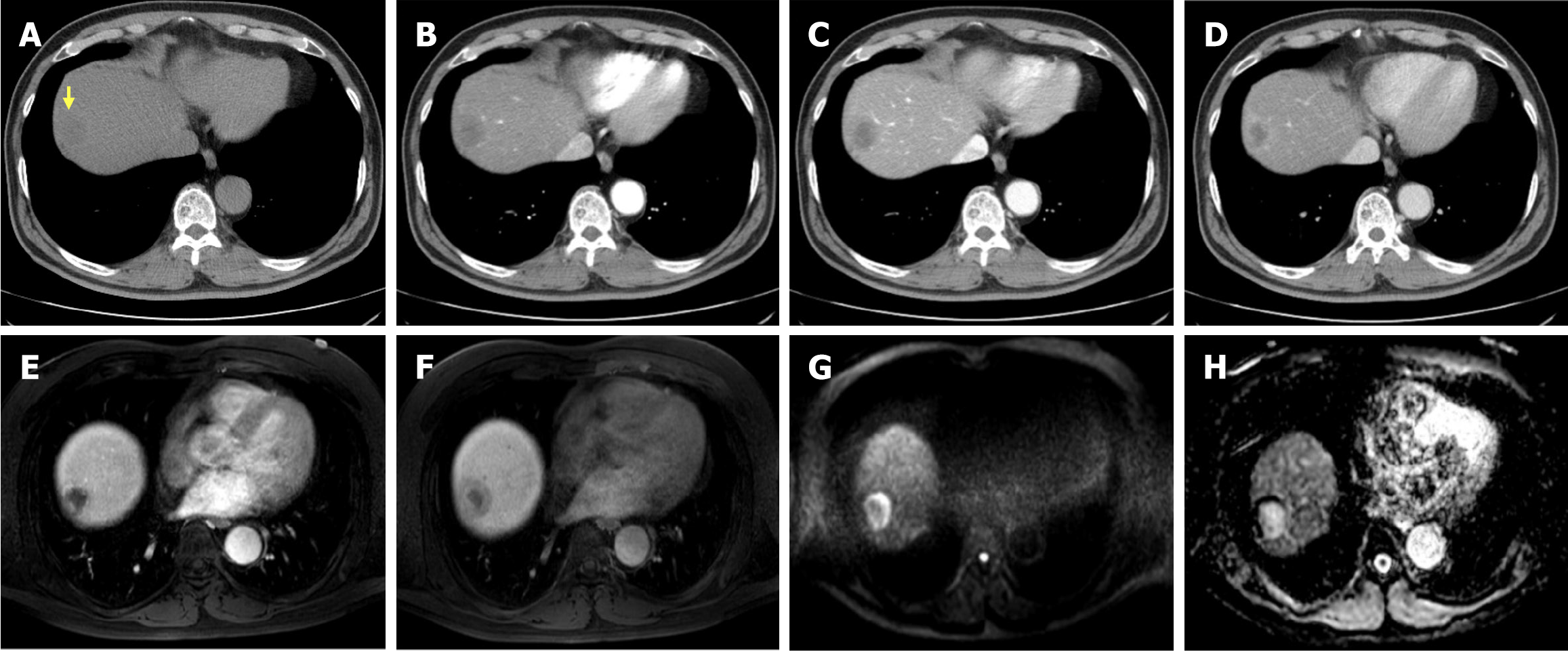
Figure 1 Preoperative abdominal dynamic computed tomography and liver magnetic resonance imaging findings.
A and B: A low-attenuation nodule (yellow arrow) in the right hepatic dome subcapsular area was observed on pre-contrast imaging, showing peripheral rim enhancement in the arterial phase (B). C and D: In the portal phase (C), the nodule demonstrates progressive centripetal enhancement, and the delayed phase (D) shows a typical centripetal enhancement pattern. E and F: Axial T1-weighted arterial-(E) and hepatobiliary-phase (F) images show peripheral enhancement with gradual centripetal filling of contrast media. G and H: Diffusion-weighted imaging with a b-value of 1000 (G) demonstrates marked peripheral high signal intensity, while the apparent diffusion coefficient map (H) reveals high signal intensity in the core of the lesion.
- Citation: Shin SH, Koh YS, Song S. Hepatic epithelioid hemangioendothelioma managed with minimally invasive surgery: A case report. World J Clin Cases 2025; 13(22): 104924
- URL: https://www.wjgnet.com/2307-8960/full/v13/i22/104924.htm
- DOI: https://dx.doi.org/10.12998/wjcc.v13.i22.104924


