Copyright
©The Author(s) 2024.
World J Clin Cases. Mar 6, 2024; 12(7): 1346-1355
Published online Mar 6, 2024. doi: 10.12998/wjcc.v12.i7.1346
Published online Mar 6, 2024. doi: 10.12998/wjcc.v12.i7.1346
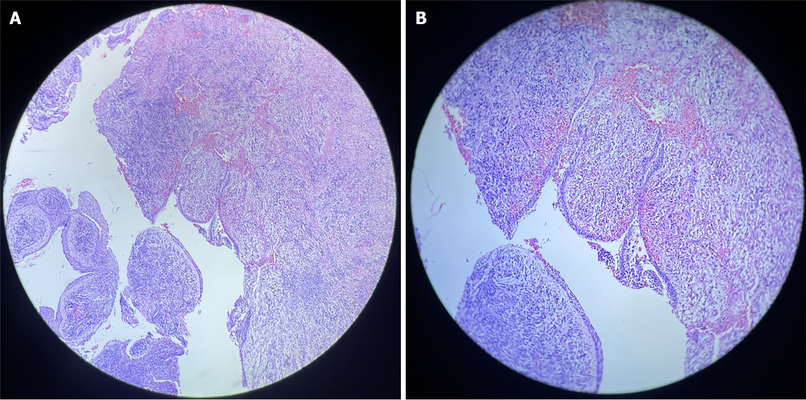
Figure 5 Histological examination.
A: Hematoxylin and eosin-stained histological sections exhibiting cavity lined by non-keratinized odontogenic epithelium with intracapsular projections beneath [Histological examination (H&E); × 20]; B: Photomicrograph showing intense, mixed inflammatory infiltrates and foamy macrophages in the fibrous capsule (H&E; × 40).
- Citation: Gómez Mireles JC, Martínez Carrillo EK, Alcalá Barbosa K, Gutiérrez Cortés E, González Ramos J, González Gómez LA, Bayardo González RA, Lomelí Martínez SM. Microsurgical management of radicular cyst using guided tissue regeneration technique: A case report. World J Clin Cases 2024; 12(7): 1346-1355
- URL: https://www.wjgnet.com/2307-8960/full/v12/i7/1346.htm
- DOI: https://dx.doi.org/10.12998/wjcc.v12.i7.1346


