Copyright
©The Author(s) 2024.
World J Clin Cases. Mar 6, 2024; 12(7): 1346-1355
Published online Mar 6, 2024. doi: 10.12998/wjcc.v12.i7.1346
Published online Mar 6, 2024. doi: 10.12998/wjcc.v12.i7.1346
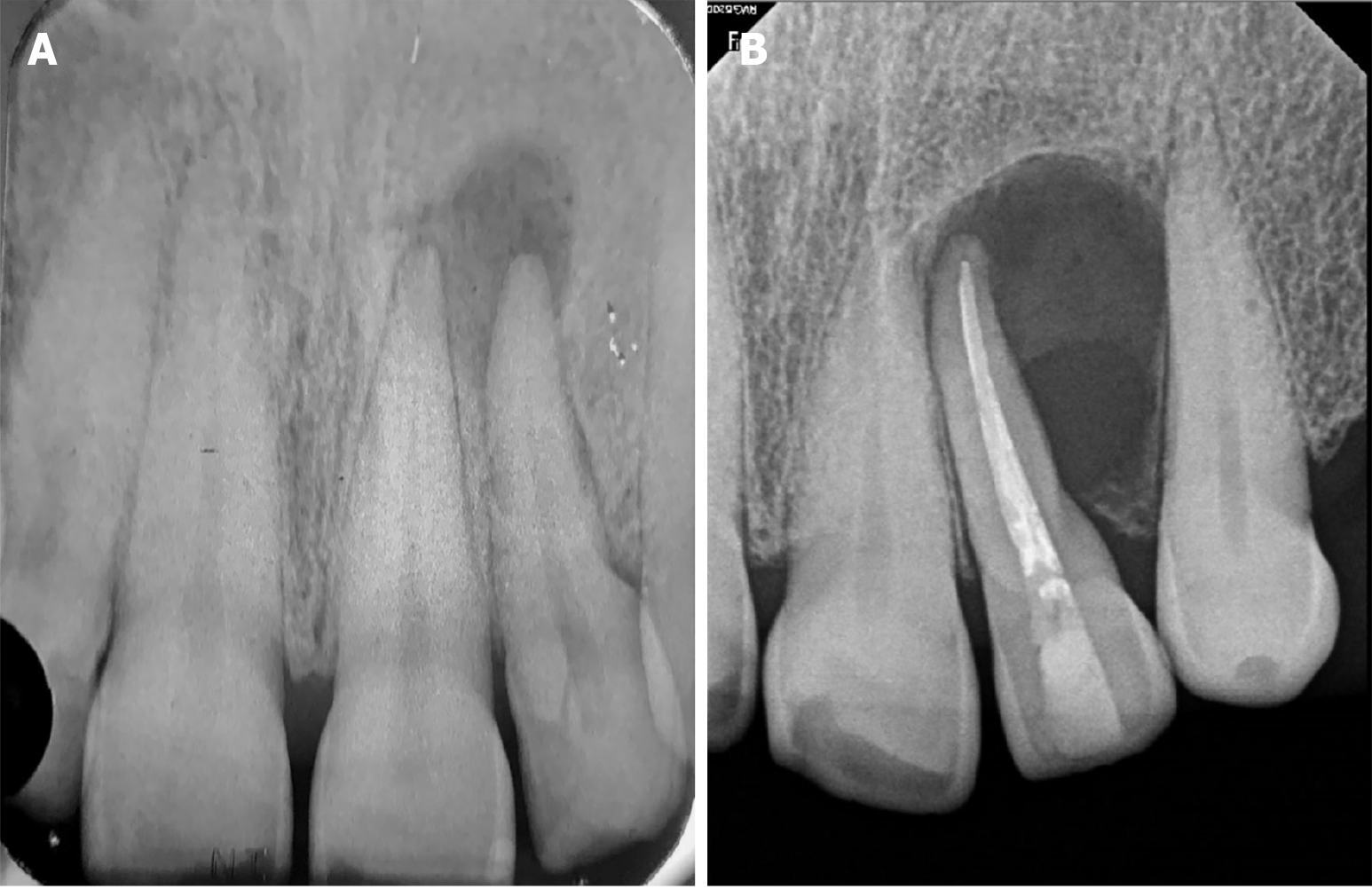
Figure 2 Preoperative periapical radiograph.
A: X-ray prior to root canal treatment, 6 years ago; B: Current initial radiograph showing a well-defined and circumscribed radiolucent area at the apex of tooth 22.
- Citation: Gómez Mireles JC, Martínez Carrillo EK, Alcalá Barbosa K, Gutiérrez Cortés E, González Ramos J, González Gómez LA, Bayardo González RA, Lomelí Martínez SM. Microsurgical management of radicular cyst using guided tissue regeneration technique: A case report. World J Clin Cases 2024; 12(7): 1346-1355
- URL: https://www.wjgnet.com/2307-8960/full/v12/i7/1346.htm
- DOI: https://dx.doi.org/10.12998/wjcc.v12.i7.1346


