Copyright
©The Author(s) 2023.
World J Clin Cases. Feb 26, 2023; 11(6): 1393-1402
Published online Feb 26, 2023. doi: 10.12998/wjcc.v11.i6.1393
Published online Feb 26, 2023. doi: 10.12998/wjcc.v11.i6.1393
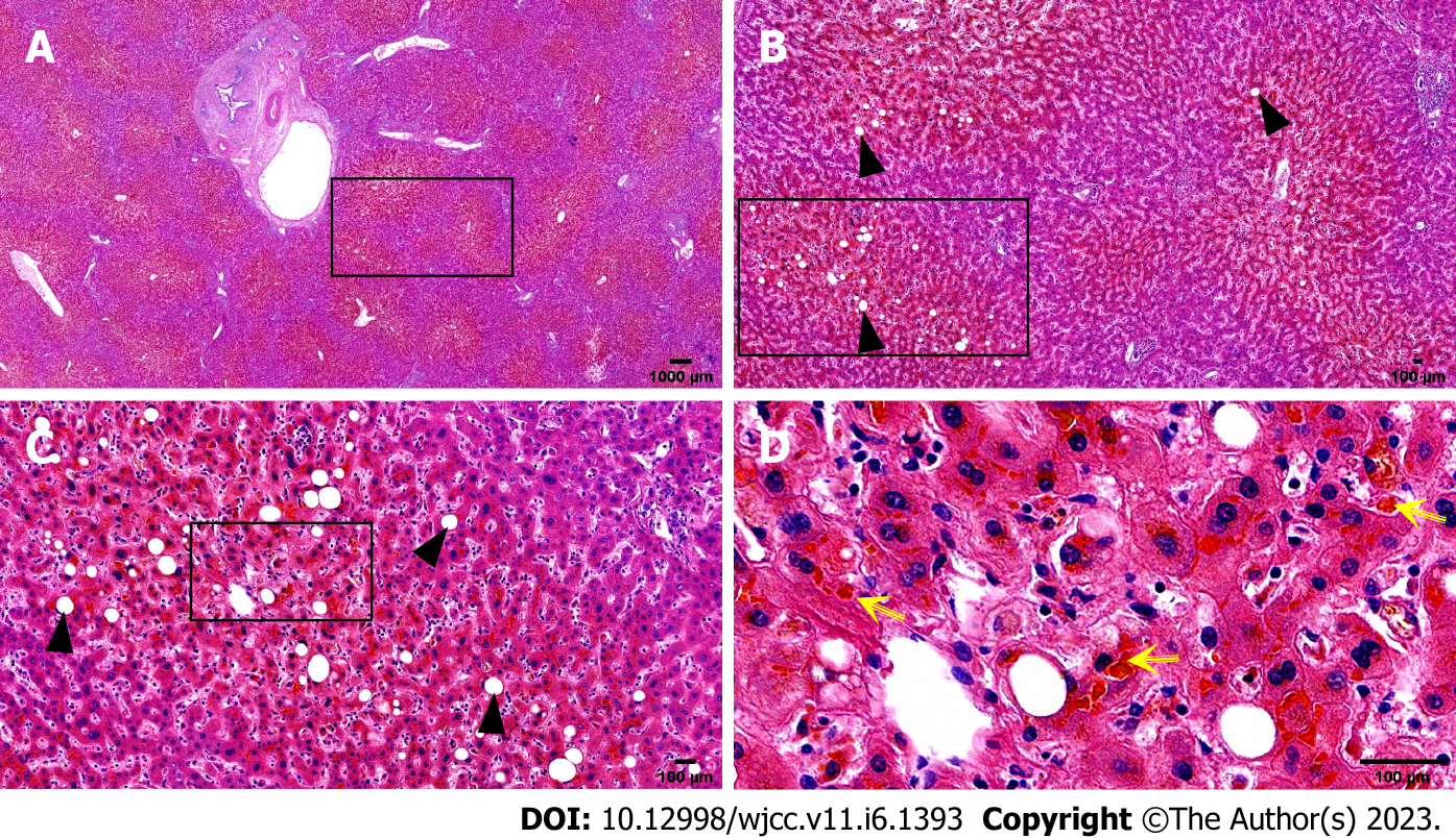
Figure 4 Histopathological biopsy of the liver.
The portal area was enlarged to varying degrees, with hyperplasia of fibrous tissue, infiltration of a moderate amount of lymphocytes and a small number of plasma cells, minor bile duct damage, fibrosis around a small number of small bile ducts, and mild bile duct hyperplasia, mild edema of hepatocytes, a few balloon-like degenerated hepatocytes, a few glycogenuclear hepatocytes, bulicular and microbulicular steatosis of hepatocytes (hepatic steatosis cells < 5%), irregular distribution, rare punctate necrosis. Some hepatocytes cholestatic pigment granules can be seen, and some bile ducts are dilated and cholestatic. Yellow arrow: Cholestatic pigment particles; Black triangle: Hepatocyte adipose change. A: Hematoxylin-eosin (HE) (× 1); B: HE (× 4); C: HE (× 10); D: HE (× 40).
- Citation: Jiang JL, Liu X, Pan ZQ, Jiang XL, Shi JH, Chen Y, Yi Y, Zhong WW, Liu KY, He YH. Postoperative jaundice related to UGT1A1 and ABCB11 gene mutations: A case report and literature review. World J Clin Cases 2023; 11(6): 1393-1402
- URL: https://www.wjgnet.com/2307-8960/full/v11/i6/1393.htm
- DOI: https://dx.doi.org/10.12998/wjcc.v11.i6.1393


