Copyright
©The Author(s) 2023.
World J Clin Cases. Feb 26, 2023; 11(6): 1393-1402
Published online Feb 26, 2023. doi: 10.12998/wjcc.v11.i6.1393
Published online Feb 26, 2023. doi: 10.12998/wjcc.v11.i6.1393
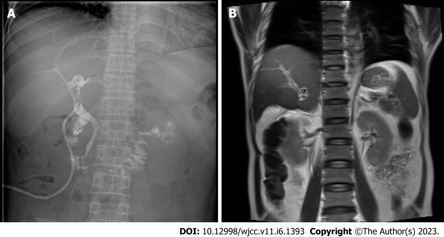
Figure 3 T-tube and magnetic resonance cholangiopancreatography images.
A: T-tube angiography: the left and right hepatic ducts were not visualized, the bile duct was rigid, and the lower end of the common bile duct was unobstructed; B: Magnetic resonance cholangiopancreatography: The intrahepatic bile duct in the upper right posterior lobe of the liver was dilated, and multiple nodular short T2 signals were seen in the lumen. Gallbladder not shown.
- Citation: Jiang JL, Liu X, Pan ZQ, Jiang XL, Shi JH, Chen Y, Yi Y, Zhong WW, Liu KY, He YH. Postoperative jaundice related to UGT1A1 and ABCB11 gene mutations: A case report and literature review. World J Clin Cases 2023; 11(6): 1393-1402
- URL: https://www.wjgnet.com/2307-8960/full/v11/i6/1393.htm
- DOI: https://dx.doi.org/10.12998/wjcc.v11.i6.1393


