Copyright
©The Author(s) 2023.
World J Clin Cases. Feb 26, 2023; 11(6): 1393-1402
Published online Feb 26, 2023. doi: 10.12998/wjcc.v11.i6.1393
Published online Feb 26, 2023. doi: 10.12998/wjcc.v11.i6.1393
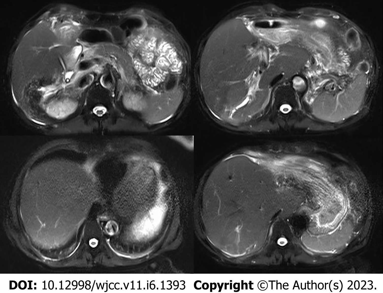
Figure 2 Postoperative magnetic resonance imaging of the upper abdomen.
Enlarged and deformed liver with uneven signals in the liver parenchyma and dilated intrahepatic bile duct in the upper right posterior lobe of the liver. Multiple nodular short T2 signal in the dilated and rigid bile duct with increased T2 weighted imaging signal in the surrounding liver parenchyma. The portal vein is widened with a size of 1.4 cm.
- Citation: Jiang JL, Liu X, Pan ZQ, Jiang XL, Shi JH, Chen Y, Yi Y, Zhong WW, Liu KY, He YH. Postoperative jaundice related to UGT1A1 and ABCB11 gene mutations: A case report and literature review. World J Clin Cases 2023; 11(6): 1393-1402
- URL: https://www.wjgnet.com/2307-8960/full/v11/i6/1393.htm
- DOI: https://dx.doi.org/10.12998/wjcc.v11.i6.1393


