Copyright
©The Author(s) 2023.
World J Clin Cases. Feb 26, 2023; 11(6): 1393-1402
Published online Feb 26, 2023. doi: 10.12998/wjcc.v11.i6.1393
Published online Feb 26, 2023. doi: 10.12998/wjcc.v11.i6.1393
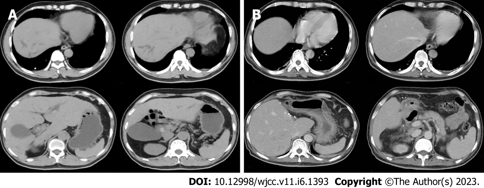
Figure 1 Computed tomography image.
A: The preoperative computed tomography (CT) image of the upper abdomen of the patient: Normal liver parenchyma with dilated intrahepatic as well as common bile duct (about 18 mm) and enlarged gallbladder without wall-thickening; B: Postoperative CT image of the upper abdomen: the enlarged and deformed remnant liver with dilated intrahepatic bile duct in the upper segment of the right posterior; the portal vein is widened, the widest diameter is about 1.4 cm. The spleen was normal in size and shape.
- Citation: Jiang JL, Liu X, Pan ZQ, Jiang XL, Shi JH, Chen Y, Yi Y, Zhong WW, Liu KY, He YH. Postoperative jaundice related to UGT1A1 and ABCB11 gene mutations: A case report and literature review. World J Clin Cases 2023; 11(6): 1393-1402
- URL: https://www.wjgnet.com/2307-8960/full/v11/i6/1393.htm
- DOI: https://dx.doi.org/10.12998/wjcc.v11.i6.1393


