Copyright
©The Author(s) 2023.
World J Clin Cases. Feb 6, 2023; 11(4): 866-873
Published online Feb 6, 2023. doi: 10.12998/wjcc.v11.i4.866
Published online Feb 6, 2023. doi: 10.12998/wjcc.v11.i4.866
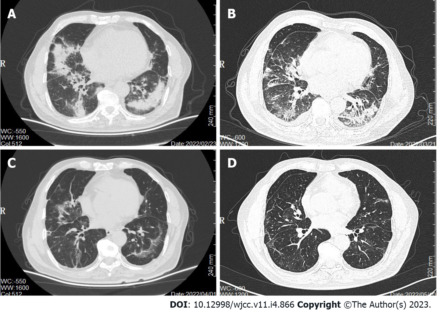
Figure 2 Chronological computed tomography of the chest demonstrating changes in the lungs.
A: Multiple patches and ground glass shadows in both lungs and scattered soft tissue nodules of different sizes in both lungs in February 2022; B: There was no significant change in pulmonary inflammation after empirical anti-infective treatment in March 2022; C: Pulmonary infection was significantly improved and resolved in both lungs in April 2022; D: Pulmonary infection almost completely disappeared in May 2022.
- Citation: Cheng QW, Shen HL, Dong ZH, Zhang QQ, Wang YF, Yan J, Wang YS, Zhang NG. Pneumocystis jirovecii diagnosed by next-generation sequencing of bronchoscopic alveolar lavage fluid: A case report and review of literature. World J Clin Cases 2023; 11(4): 866-873
- URL: https://www.wjgnet.com/2307-8960/full/v11/i4/866.htm
- DOI: https://dx.doi.org/10.12998/wjcc.v11.i4.866


