Copyright
©The Author(s) 2023.
World J Clin Cases. Nov 26, 2023; 11(33): 8078-8083
Published online Nov 26, 2023. doi: 10.12998/wjcc.v11.i33.8078
Published online Nov 26, 2023. doi: 10.12998/wjcc.v11.i33.8078
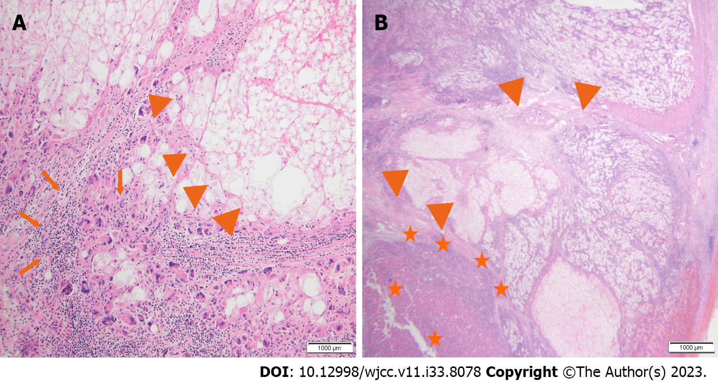
Figure 3 Postoperative histopathological findings.
A and B: A significant portion of the liver parenchyma has undergone necrosis, showing abscess-like features (arrowhead), inflammatory cell infiltration (arrow) and hepatocellular carcinoma tumor cells (star) in the remaining liver parenchyma.
- Citation: Ryou SH, Shin HD, Kim SB. Hepatocellular carcinoma presenting as organized liver abscess: A case report. World J Clin Cases 2023; 11(33): 8078-8083
- URL: https://www.wjgnet.com/2307-8960/full/v11/i33/8078.htm
- DOI: https://dx.doi.org/10.12998/wjcc.v11.i33.8078


