Copyright
©The Author(s) 2023.
World J Clin Cases. Jan 26, 2023; 11(3): 645-654
Published online Jan 26, 2023. doi: 10.12998/wjcc.v11.i3.645
Published online Jan 26, 2023. doi: 10.12998/wjcc.v11.i3.645
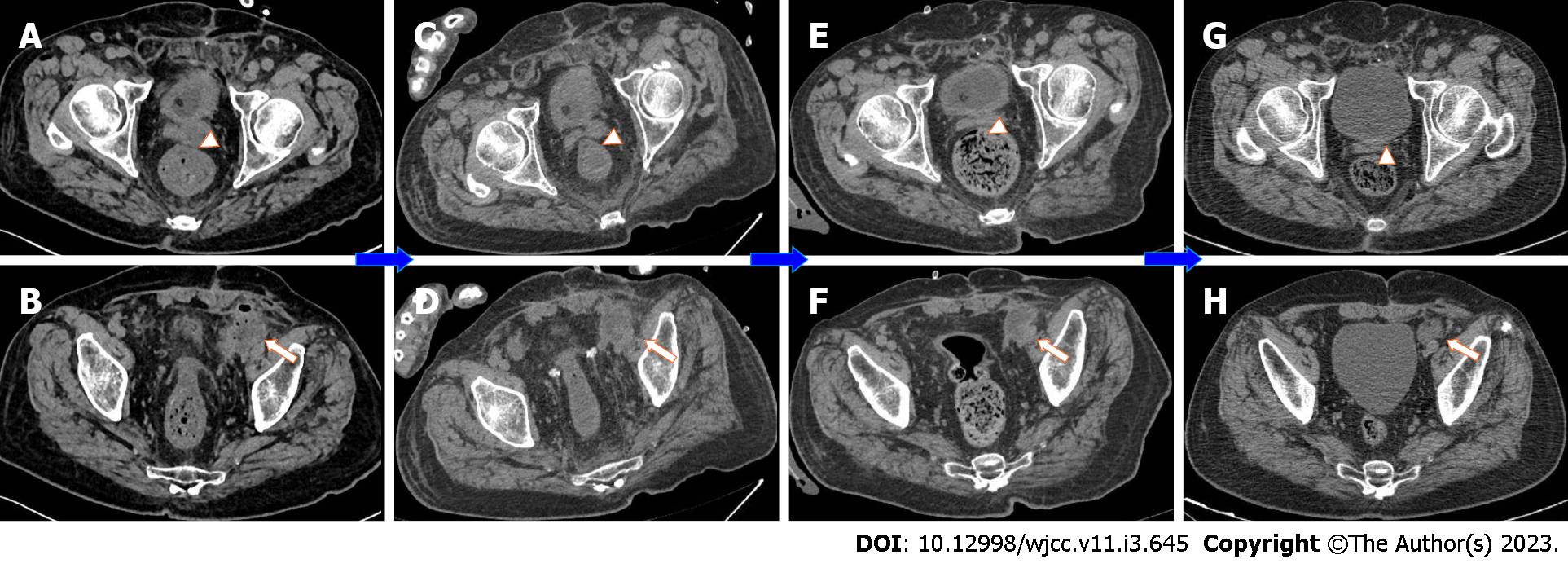
Figure 2 Computed tomography results of the patient throughout the diagnosis and treatment processes.
A-D: The results of pelvic computed tomography examinations on the 2nd day after admission, on the 10th day after surgery, at 1 mo after discharge, and at 3 mo after discharge; E-H: The primary focus of the left seminal vesicle abscess and the secondary focus of the extraperitoneal fascial abscess were gradually controlled and narrowed after treatment and outpatient and emergency follow-up. The white arrowheads show the left seminal vesicle. The white arrows show the pelvic abscess (secondary focus).
- Citation: Li K, Liu NB, Liu JX, Chen QN, Shi BM. Acute diffuse peritonitis secondary to a seminal vesicle abscess: A case report. World J Clin Cases 2023; 11(3): 645-654
- URL: https://www.wjgnet.com/2307-8960/full/v11/i3/645.htm
- DOI: https://dx.doi.org/10.12998/wjcc.v11.i3.645


