Copyright
©The Author(s) 2023.
World J Clin Cases. Oct 16, 2023; 11(29): 7234-7241
Published online Oct 16, 2023. doi: 10.12998/wjcc.v11.i29.7234
Published online Oct 16, 2023. doi: 10.12998/wjcc.v11.i29.7234
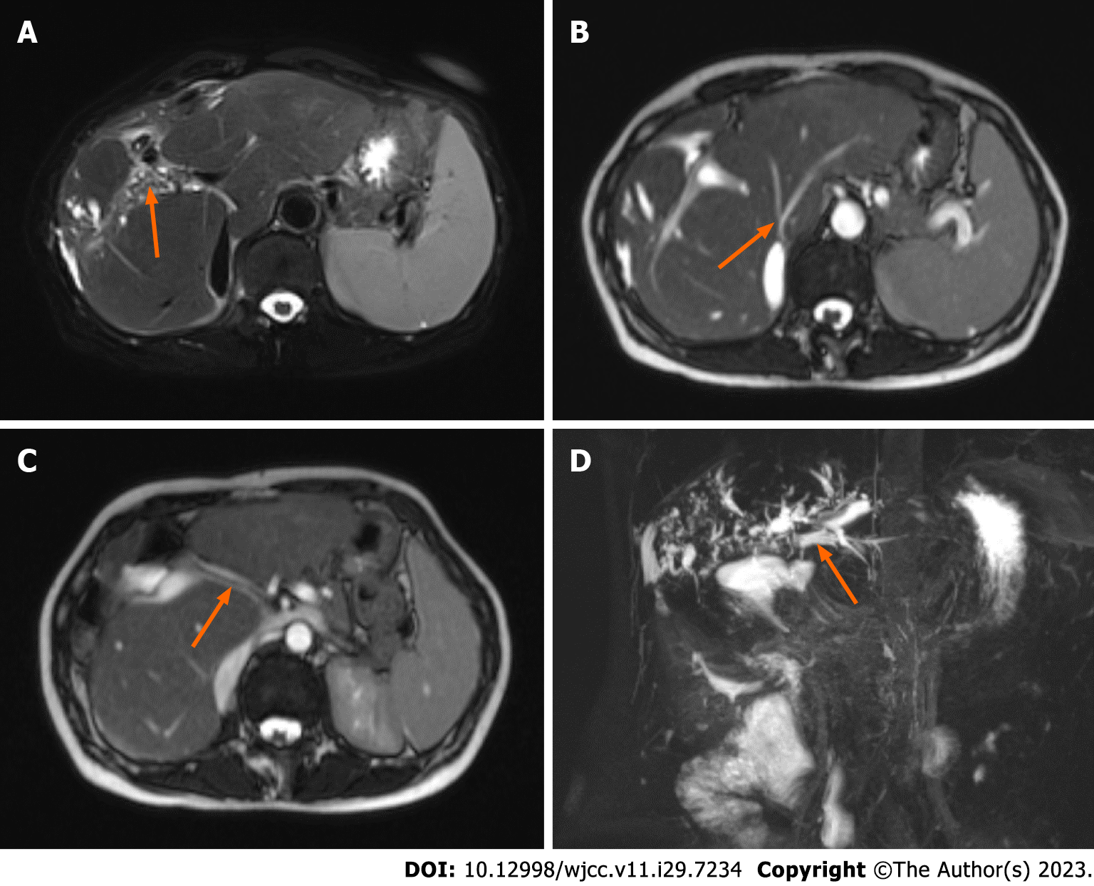
Figure 2 Abdominal computed tomography image.
A: Multiple stones in the intrahepatic bile duct; B: The hepatic veins draining back into the inferior vena cava were narrowed; C: The main portal vein runs through the middle of the liver lobe; D: Magnetic resonance cholangiopancreatography showed dilatation of intrahepatic bile ducts, indicated by the orange arrow and did not show bilioenteric anastomosis.
- Citation: Liang SY, Lu JG, Wang ZD. Imaging misdiagnosis and clinical analysis of significant hepatic atrophy after bilioenteric anastomosis: A case report. World J Clin Cases 2023; 11(29): 7234-7241
- URL: https://www.wjgnet.com/2307-8960/full/v11/i29/7234.htm
- DOI: https://dx.doi.org/10.12998/wjcc.v11.i29.7234


