Copyright
©The Author(s) 2023.
World J Clin Cases. Sep 16, 2023; 11(26): 6213-6222
Published online Sep 16, 2023. doi: 10.12998/wjcc.v11.i26.6213
Published online Sep 16, 2023. doi: 10.12998/wjcc.v11.i26.6213
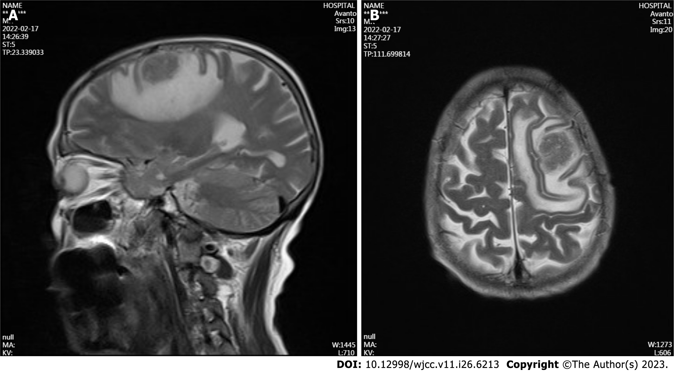
Figure 4 Brain magnetic resonance imaging in February 2022.
The mass in the left frontal lobe was approximately 2.6 cm in diameter, with a distinct band of surrounding edema. A: Median sagittal section of brain magnetic resonance imaging (MRI); B: Transverse section of brain MRI.
- Citation: Weng XT, Lin WL, Pan QM, Chen TF, Li SY, Gu CM. Aggressive variant prostate cancer: A case report and literature review. World J Clin Cases 2023; 11(26): 6213-6222
- URL: https://www.wjgnet.com/2307-8960/full/v11/i26/6213.htm
- DOI: https://dx.doi.org/10.12998/wjcc.v11.i26.6213


