
















©The Author(s) 2023.
World J Clin Cases. Jul 16, 2023; 11(20): 4903-4911
Published online Jul 16, 2023. doi: 10.12998/wjcc.v11.i20.4903
Published online Jul 16, 2023. doi: 10.12998/wjcc.v11.i20.4903
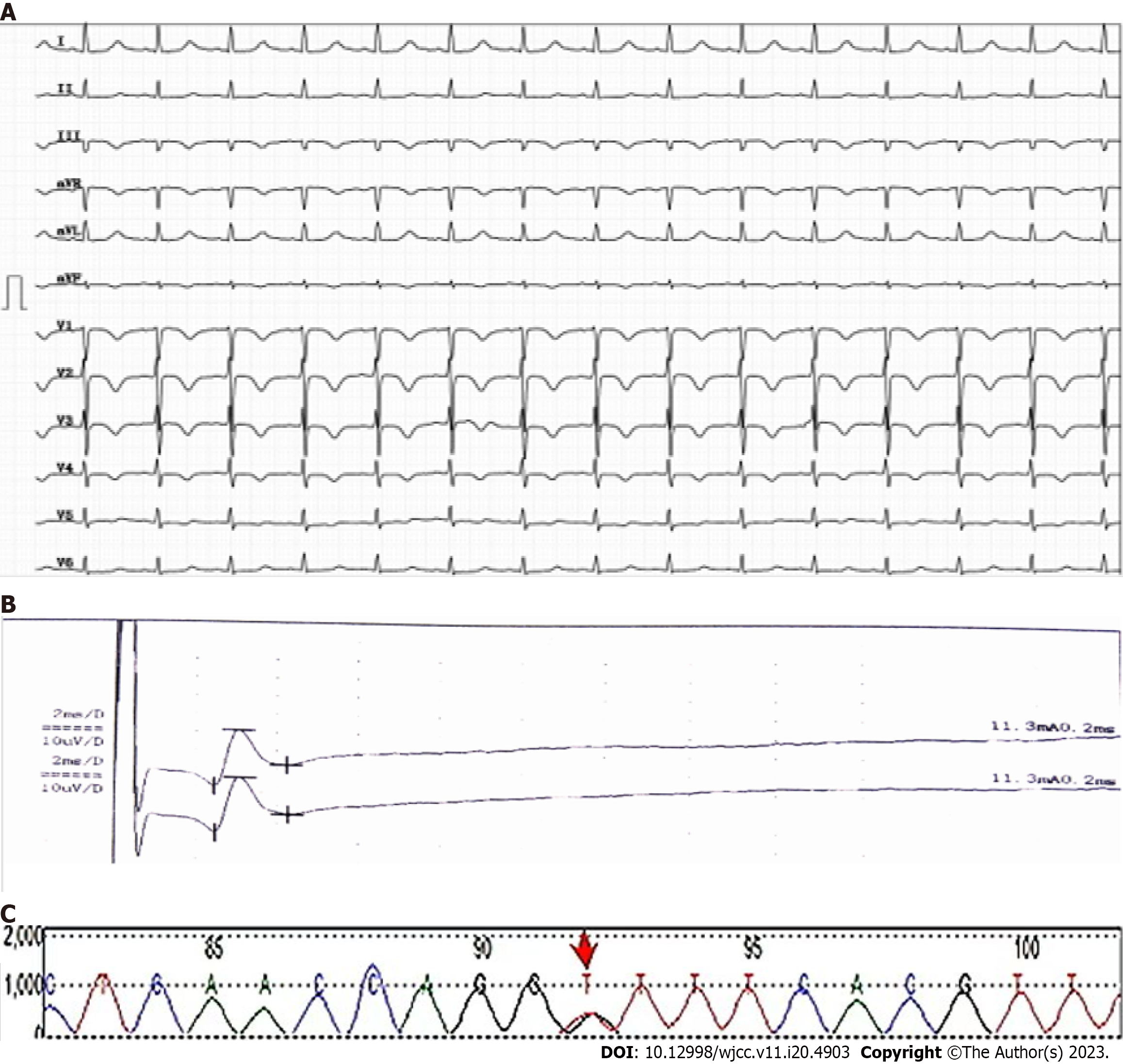
Figure 3 Electrocardiogram, electromyography, and ATP-binding cassette, subfamily B gene mutation sites.
A: Electrocardiogram from January 2022. Note: The electrocardiogram shows sinus tachycardia, heart rate 124 bpm, left ventricular high voltage T wave changes (V1-V4 low level, inversion), and QT interval prolongation; B: Electromyography from January 2022. Note: The electromyography showed that the conduction velocity of the bilateral superficial peroneal nerve was slightly slowed and the amplitude was reduced; C: The ATP-binding cassette, subfamily B (ABCB4) c.2318 G>T heterozygous mutation. Note: The red arrow shows a G to T transformation of the 2318 base on chromosome 7, resulting in the transformation of codon 773 from Gly to Val and the encoding product from glycine to valine.
- Citation: Cheng N, Qin YJ, Zhang Q, Li H. ABCB4 gene mutation-associated cirrhosis with systemic amyloidosis: A case report. World J Clin Cases 2023; 11(20): 4903-4911
- URL: https://www.wjgnet.com/2307-8960/full/v11/i20/4903.htm
- DOI: https://dx.doi.org/10.12998/wjcc.v11.i20.4903