Copyright
©The Author(s) 2023.
World J Clin Cases. Jul 16, 2023; 11(20): 4903-4911
Published online Jul 16, 2023. doi: 10.12998/wjcc.v11.i20.4903
Published online Jul 16, 2023. doi: 10.12998/wjcc.v11.i20.4903
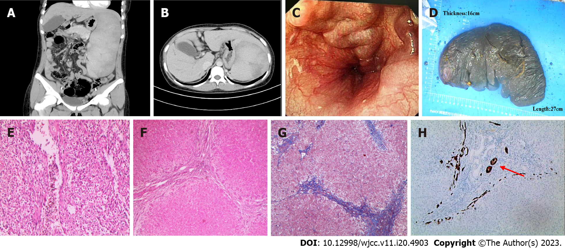
Figure 1 Epigastrium enhanced computed tomography, gastroscopy, postoperative spleen appearance, pathological image.
A: On November 2018, computed tomography (CT) of upper abdomen enhanced sagittal plane; B: CT enhanced coronal plane of upper abdomen in November 2018; C: Gastroscopy revealed esophageal varices; D: Spleen appearance (27 cm × 16 cm); E: Spleen stained with hematoxylin and eosin (H&E) × 100; F: H&E staining of liver tissue × 100; G: Masson staining of liver tissue (original magnification × 100); H: CK7 staining of liver tissue (original magnification × 100).
- Citation: Cheng N, Qin YJ, Zhang Q, Li H. ABCB4 gene mutation-associated cirrhosis with systemic amyloidosis: A case report. World J Clin Cases 2023; 11(20): 4903-4911
- URL: https://www.wjgnet.com/2307-8960/full/v11/i20/4903.htm
- DOI: https://dx.doi.org/10.12998/wjcc.v11.i20.4903


