Copyright
©The Author(s) 2023.
World J Clin Cases. Jun 16, 2023; 11(17): 4159-4167
Published online Jun 16, 2023. doi: 10.12998/wjcc.v11.i17.4159
Published online Jun 16, 2023. doi: 10.12998/wjcc.v11.i17.4159
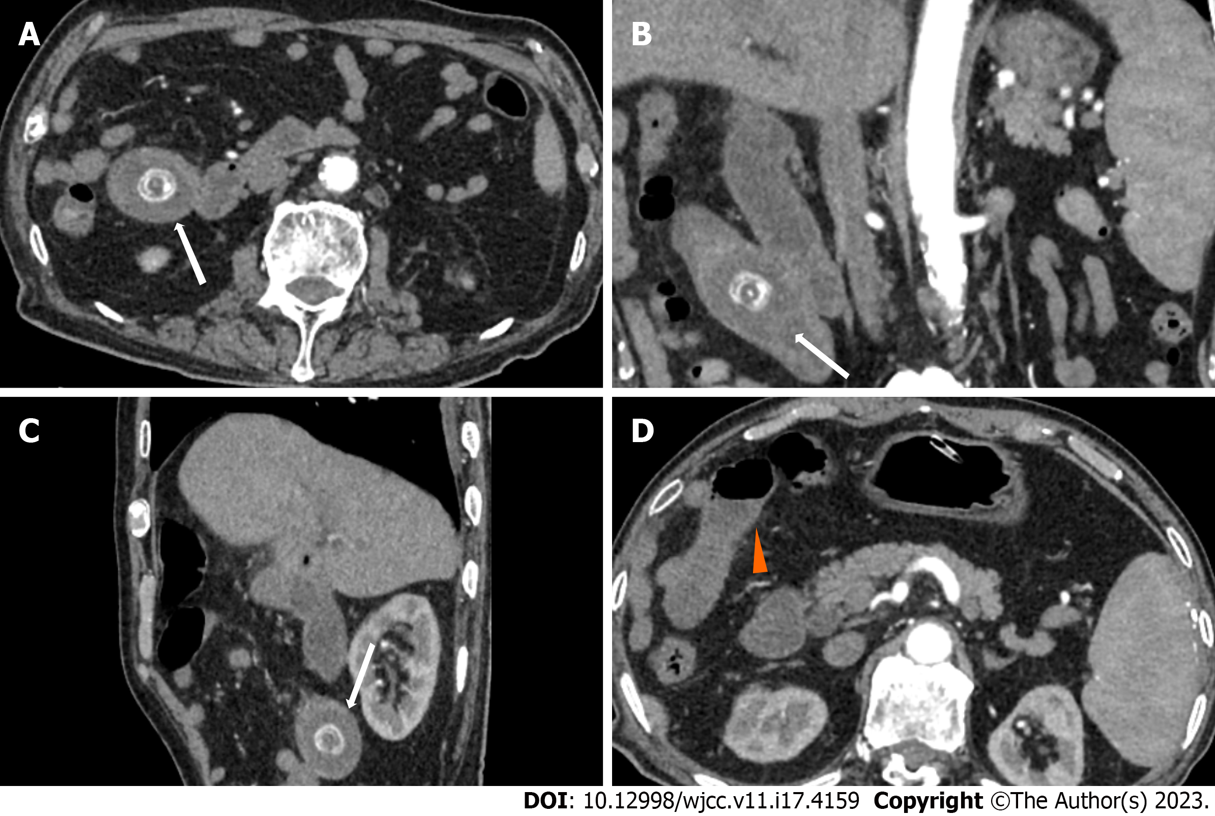
Figure 3 Abdominal contrast enhanced computed tomography of the jejunal gallstone ileus.
A-C: A stone was shown in the upper jejunum (white arrows); D: Proximal intestinal effusion and dilatation (orange arrow).
- Citation: Fan WJ, Liu M, Feng XX. Endoscopic and surgical treatment of jejunal gallstone ileus caused by cholecystoduodenal fistula: A case report. World J Clin Cases 2023; 11(17): 4159-4167
- URL: https://www.wjgnet.com/2307-8960/full/v11/i17/4159.htm
- DOI: https://dx.doi.org/10.12998/wjcc.v11.i17.4159


