
















©The Author(s) 2023.
World J Clin Cases. May 26, 2023; 11(15): 3625-3630
Published online May 26, 2023. doi: 10.12998/wjcc.v11.i15.3625
Published online May 26, 2023. doi: 10.12998/wjcc.v11.i15.3625
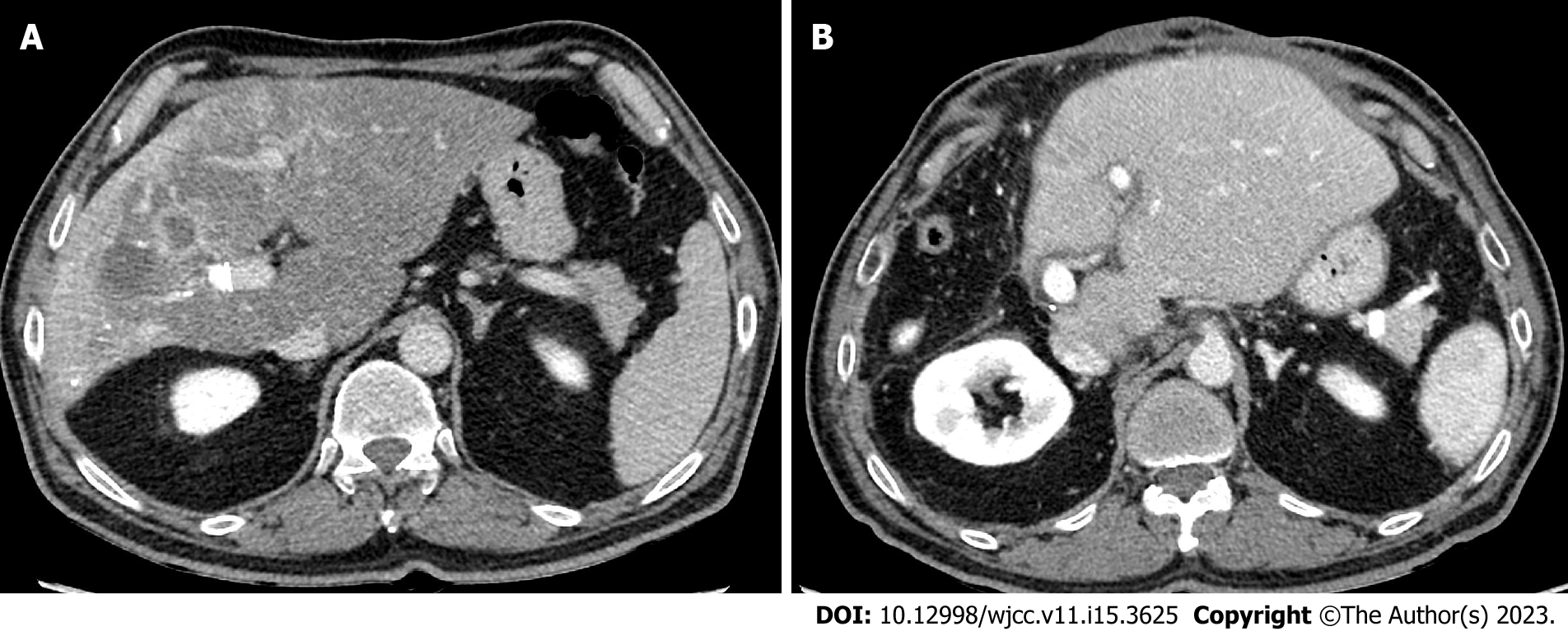
Figure 3 Contrast enhanced computed tomography of the liver.
A: Axial computed tomography (CT) showed post portal vein embolization shrinkage of right liver lobe and hypertrophy of left liver lobe; B: Axial CT showed post-operative hypertrophic left hepatic lobe.
- Citation: Alharbi SR, Bin Nasif M, Alwaily HB. Non-target lung embolization during portal vein embolization due to an unrecognized portosystemic venous fistula: A case report. World J Clin Cases 2023; 11(15): 3625-3630
- URL: https://www.wjgnet.com/2307-8960/full/v11/i15/3625.htm
- DOI: https://dx.doi.org/10.12998/wjcc.v11.i15.3625