Copyright
©The Author(s) 2023.
World J Clin Cases. May 16, 2023; 11(14): 3340-3350
Published online May 16, 2023. doi: 10.12998/wjcc.v11.i14.3340
Published online May 16, 2023. doi: 10.12998/wjcc.v11.i14.3340
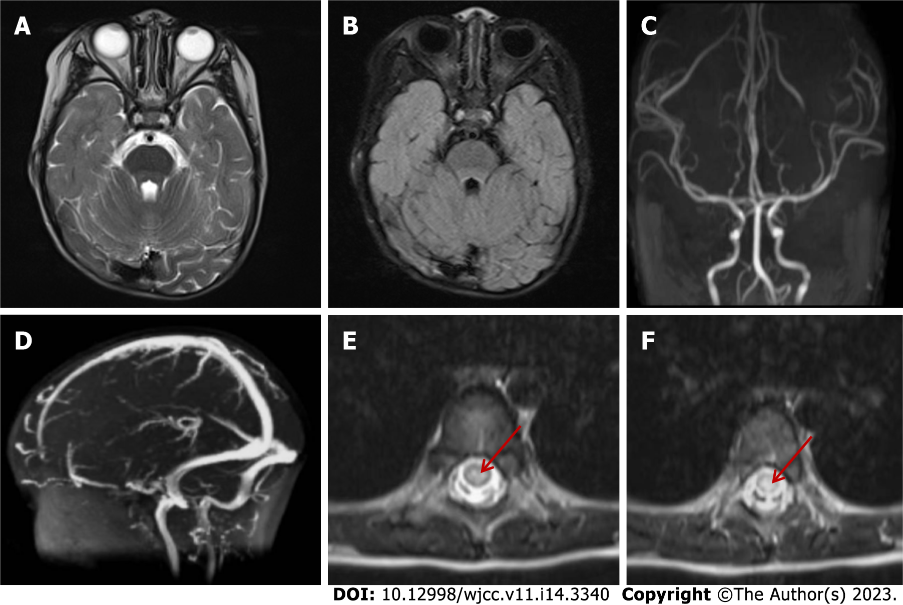
Figure 1 The imaging findings of brain and spinal cord magnetic resonance on day 2 after admission.
A and B: T2-weighted magnetic resonance imaging and fluid-attenuated inversion-recovery imaging of the head, respectively, showed no abnormal signals in the brainstem, cerebellum, and cerebral cortex; C: Magnetic resonance arterial angiography of the head was normal; D: Brain magnetic resonance venography was normal; E and F: T2 hyperintensity in T7-10 horizontal transverse section of thoracic spinal cord (indicated by red arrow).
- Citation: Ding L, Huang TT, Ying GH, Wang SY, Xu HF, Qian H, Rahman F, Lu XP, Guo H, Zheng G, Zhang G. De novo mutation of NAXE (APOAIBP)-related early-onset progressive encephalopathy with brain edema and/or leukoencephalopathy-1: A case report. World J Clin Cases 2023; 11(14): 3340-3350
- URL: https://www.wjgnet.com/2307-8960/full/v11/i14/3340.htm
- DOI: https://dx.doi.org/10.12998/wjcc.v11.i14.3340


