Copyright
©The Author(s) 2023.
World J Clin Cases. Jan 6, 2023; 11(1): 233-241
Published online Jan 6, 2023. doi: 10.12998/wjcc.v11.i1.233
Published online Jan 6, 2023. doi: 10.12998/wjcc.v11.i1.233
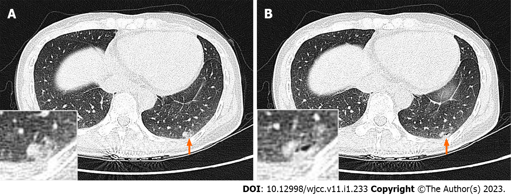
Figure 1 High-resolution computed tomography.
A: A mixed solid and ground-glass nodule (orange arrow, magnified in insert) was present in the posterior basal segment of the lower lobe of the left lung. It measures approximately 17.0 mm × 7.0 mm, was irregular in shape, and was close to the pleura; B: There was a small cystic cavity within the nodule (orange arrow, magnified in insert).
- Citation: Liu XL, Miao CF, Li M, Li P. Malignant transformation of pulmonary bronchiolar adenoma into mucinous adenocarcinoma: A case report. World J Clin Cases 2023; 11(1): 233-241
- URL: https://www.wjgnet.com/2307-8960/full/v11/i1/233.htm
- DOI: https://dx.doi.org/10.12998/wjcc.v11.i1.233


