Copyright
©The Author(s) 2023.
World J Clin Cases. Jan 6, 2023; 11(1): 177-186
Published online Jan 6, 2023. doi: 10.12998/wjcc.v11.i1.177
Published online Jan 6, 2023. doi: 10.12998/wjcc.v11.i1.177
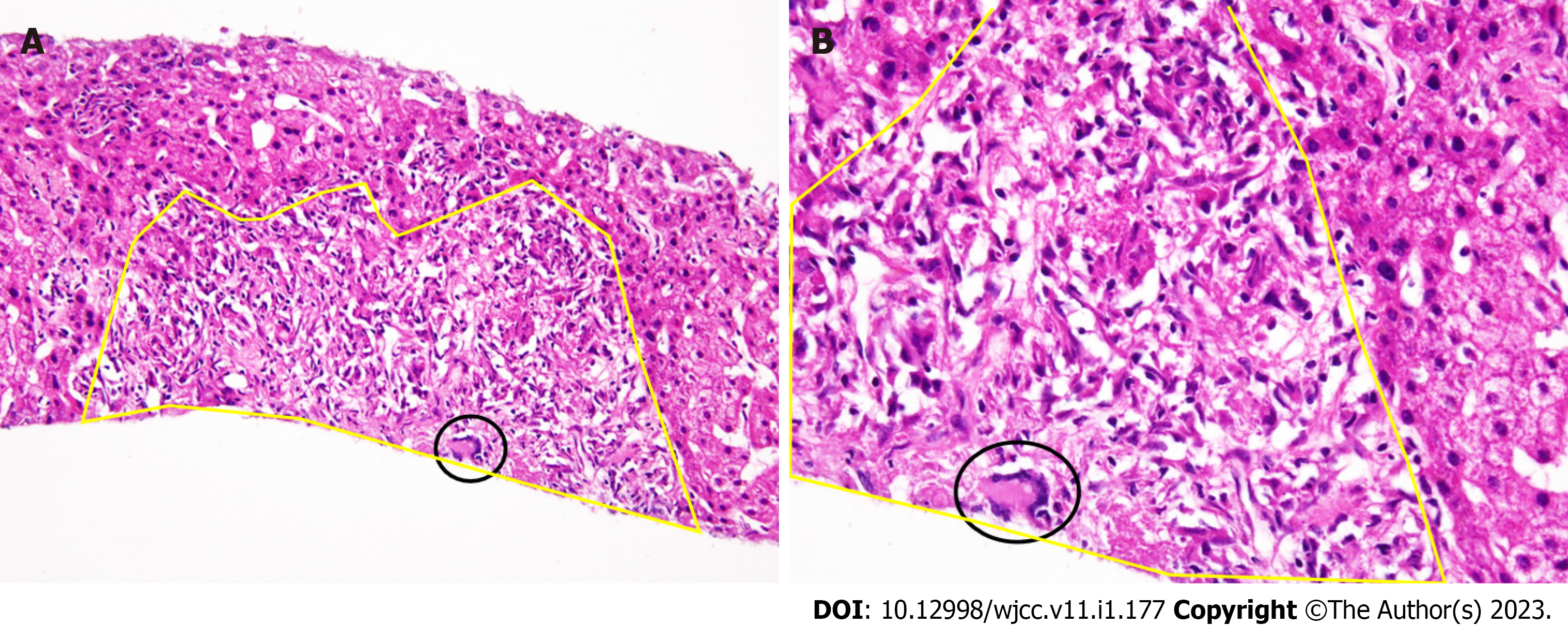
Figure 3 Histopathological findings.
A: Hematoxylin and eosin (HE) staining, low magnification Noncaseating hepatic sarcoid-like epithelioid granuloma with spindle shaped epithelioid cells (encompassed by yellow line) harboring Langhans-type multinucleated giant cells (circled black) (HE × 40); B: HE staining, high magnification of A (HE × 100).
- Citation: Kim SR, Kim SK, Fujii T, Kobayashi H, Okuda T, Hayakumo T, Nakai A, Fujii Y, Suzuki R, Sasase N, Otani A, Koma YI, Sasaki M, Kumabe T, Nakashima O. Drug-induced sarcoidosis-like reaction three months after BNT162b2 mRNA COVID-19 vaccination: A case report and review of literature. World J Clin Cases 2023; 11(1): 177-186
- URL: https://www.wjgnet.com/2307-8960/full/v11/i1/177.htm
- DOI: https://dx.doi.org/10.12998/wjcc.v11.i1.177


