Copyright
©The Author(s) 2022.
World J Clin Cases. Mar 16, 2022; 10(8): 2543-2549
Published online Mar 16, 2022. doi: 10.12998/wjcc.v10.i8.2543
Published online Mar 16, 2022. doi: 10.12998/wjcc.v10.i8.2543
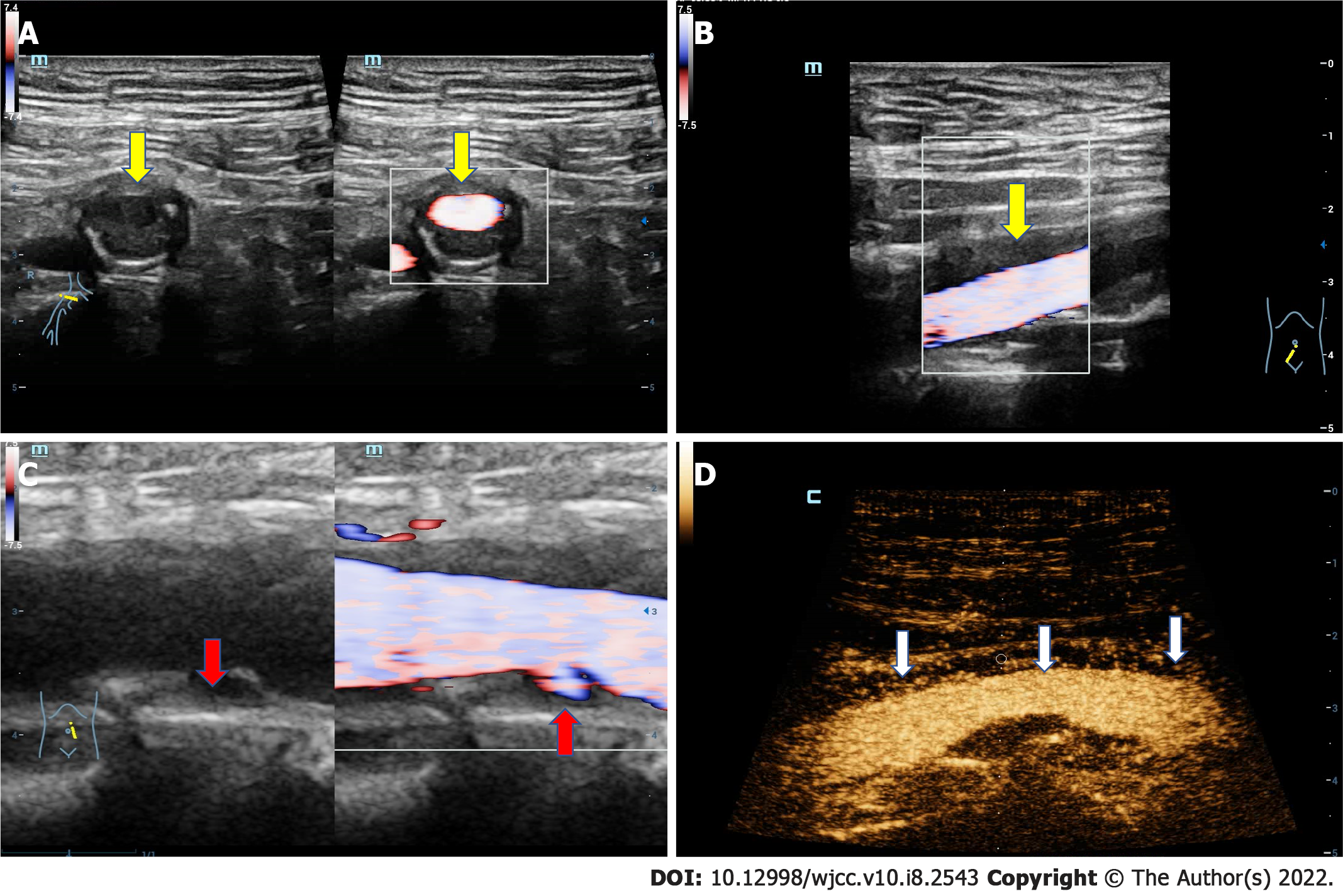
Figure 3 The ultrasound images of the artery.
A: The transverse ultrasound image of iliac artery shows that the adventitia is obviously thickened; B: The longitudinal ultrasound image of iliac artery shows that the adventitia is obviously thickened (yellow arrow); C: The longitudinal section of iliac artery, in which multiple plaques can be clearly seen on the arterial wall, and there is an ulceration in the plaque of the posterior wall (red arrow); D: Contrast-enhanced ultrasound demonstrates that extensive new blood vessels in the adventitia (white arrow).
- Citation: An YQ, Ma N, Liu Y. Immunoglobulin G4-related disease involving multiple systems: A case report. World J Clin Cases 2022; 10(8): 2543-2549
- URL: https://www.wjgnet.com/2307-8960/full/v10/i8/2543.htm
- DOI: https://dx.doi.org/10.12998/wjcc.v10.i8.2543


