
















©The Author(s) 2022.
World J Clin Cases. Mar 16, 2022; 10(8): 2543-2549
Published online Mar 16, 2022. doi: 10.12998/wjcc.v10.i8.2543
Published online Mar 16, 2022. doi: 10.12998/wjcc.v10.i8.2543
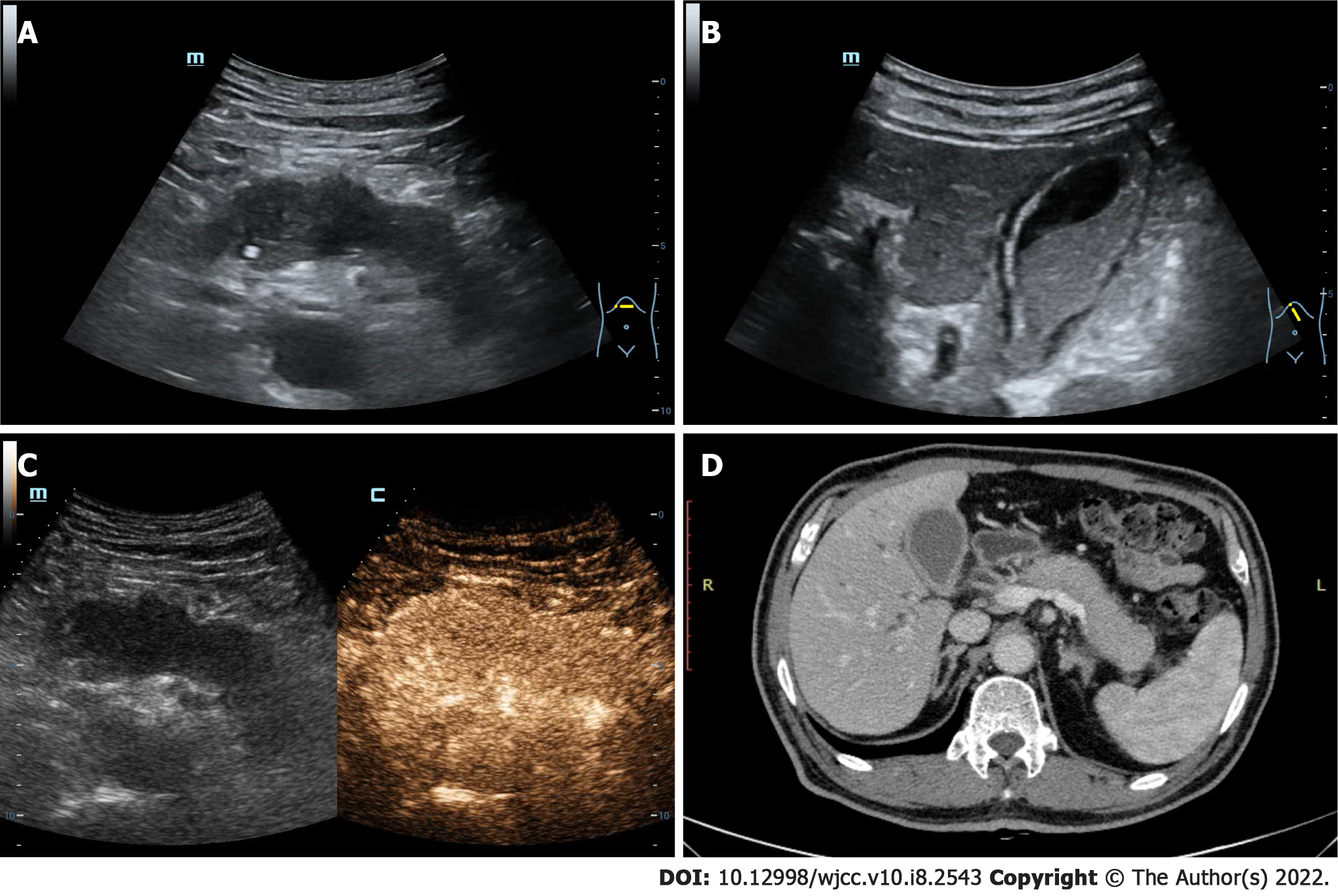
Figure 1 The ultrasound and computed tomography images of pancreas and gallbladder.
A: The pancreas is diffusely enlarged with unclear boundary and the parenchyma echo is reduced; B: The gallbladder volume is enlarged and the wall is rough, accompanying with the silt-like deposits; C: The pancreatic lesions area is uniformly enhanced in arterial phase after the intravenous ultrasound contrast-enhanced; D: Contrast-enhanced computed tomography image revealing that both the pancreas and gallbladder are enlarged and the gallbladder wall is thickened, which are consistent with the ultrasound results.
- Citation: An YQ, Ma N, Liu Y. Immunoglobulin G4-related disease involving multiple systems: A case report. World J Clin Cases 2022; 10(8): 2543-2549
- URL: https://www.wjgnet.com/2307-8960/full/v10/i8/2543.htm
- DOI: https://dx.doi.org/10.12998/wjcc.v10.i8.2543