Copyright
©The Author(s) 2022.
World J Clin Cases. Dec 16, 2022; 10(35): 13022-13027
Published online Dec 16, 2022. doi: 10.12998/wjcc.v10.i35.13022
Published online Dec 16, 2022. doi: 10.12998/wjcc.v10.i35.13022
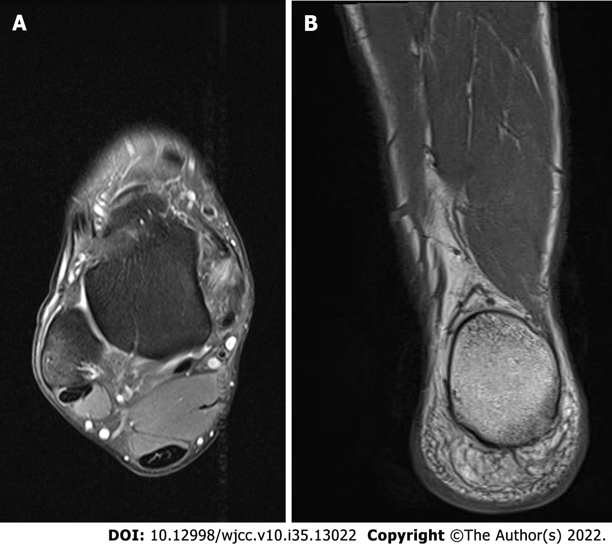
Figure 2 Magnetic resonance images of accessory soleus occupying the posterior compartment of the right ankle.
A: Accessory soleus caused bulging of the flexor hallucis longus in axial T2 fat suppressed view; B: In coronal T1-weighted view, the accessory soleus muscle was widely distributed at the medial calcaneal tuberosity.
- Citation: Woo I, Park CH, Yan H, Park JJ. Symptomatic accessory soleus muscle: A cause for exertional compartment syndrome in a young soldier: A case report. World J Clin Cases 2022; 10(35): 13022-13027
- URL: https://www.wjgnet.com/2307-8960/full/v10/i35/13022.htm
- DOI: https://dx.doi.org/10.12998/wjcc.v10.i35.13022


