Copyright
©The Author(s) 2022.
World J Clin Cases. Nov 26, 2022; 10(33): 12261-12267
Published online Nov 26, 2022. doi: 10.12998/wjcc.v10.i33.12261
Published online Nov 26, 2022. doi: 10.12998/wjcc.v10.i33.12261
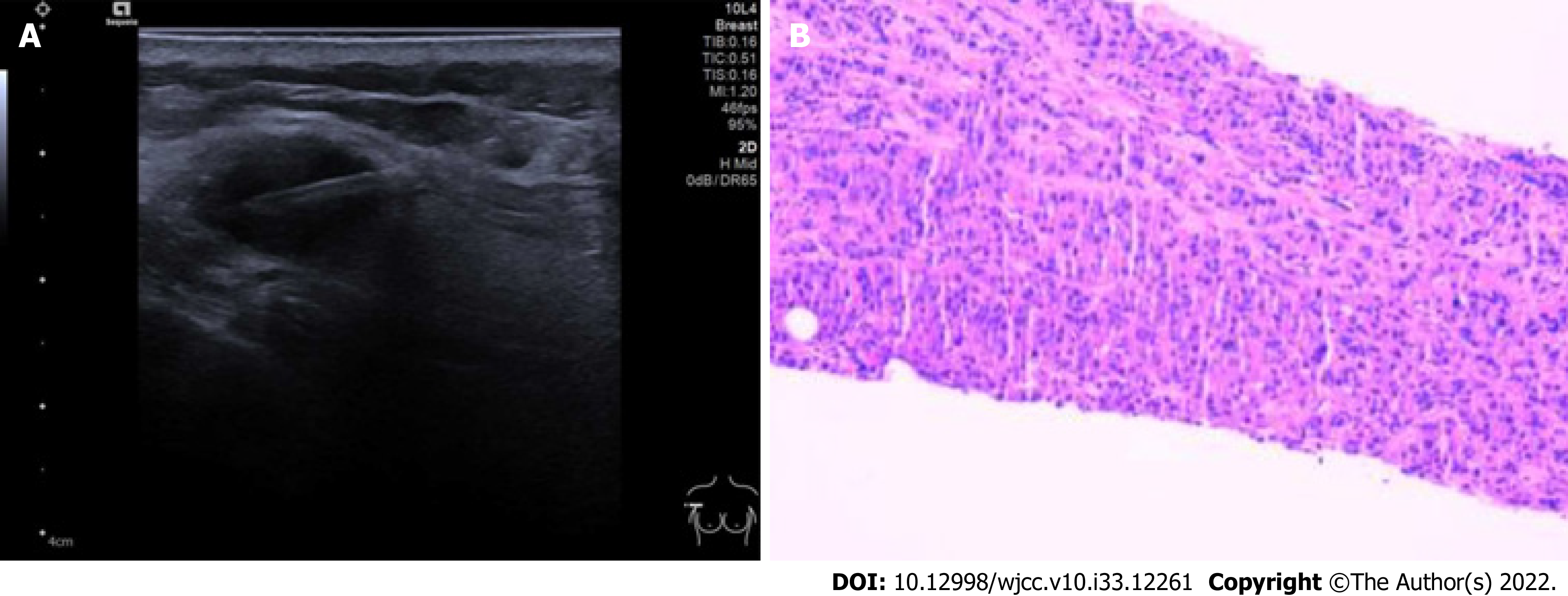
Figure 4 Brachial plexus metastasis from breast cancer.
A: The patient underwent lesion punctuation under ultrasound guidance and the lesion was sent for pathology; B: Pathology showed large areas of tumor cells in fibroblast tissue. Immunohistochemistry showed the following results: A2-1: GATA3 (+), ER (+, strong, 90%), PR (+, moderate, 10%), HER-2 (3+), Ki67 (+15%), P120 (membrane+), P63 (-), E-cadherin (+), CK5/6 (-).
- Citation: Zeng Z, Lin N, Sun LT, Chen CX. Subclavian brachial plexus metastasis from breast cancer: A case report. World J Clin Cases 2022; 10(33): 12261-12267
- URL: https://www.wjgnet.com/2307-8960/full/v10/i33/12261.htm
- DOI: https://dx.doi.org/10.12998/wjcc.v10.i33.12261


