
















©The Author(s) 2022.
World J Clin Cases. Nov 26, 2022; 10(33): 12247-12256
Published online Nov 26, 2022. doi: 10.12998/wjcc.v10.i33.12247
Published online Nov 26, 2022. doi: 10.12998/wjcc.v10.i33.12247
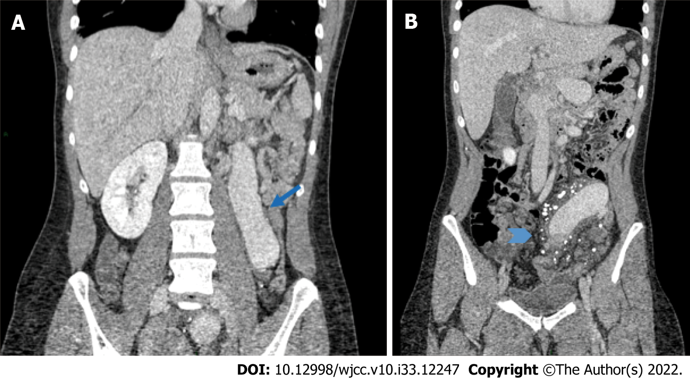
Figure 2 Abdominal computed tomography showing severe mesenteric vein dilatation.
A: Coronal reformatted contrast enhanced computed tomography showing the significant varicose dilatation of the inferior mesenteric vein (arrow); B: Striation of the fatty tissue surrounding the inferior mesenteric vein, with the presence of multiple calcified granulomas suggesting chronic calcifications of small confluent branches (arrow head).
- Citation: Azrad-Daniel S, Cupa-Galvan C, Farca-Soffer S, Perez-Zincer F, Lopez-Acosta ME. Unusual presentation of Loeys-Dietz syndrome: A case report of clinical findings and treatment challenges. World J Clin Cases 2022; 10(33): 12247-12256
- URL: https://www.wjgnet.com/2307-8960/full/v10/i33/12247.htm
- DOI: https://dx.doi.org/10.12998/wjcc.v10.i33.12247