Copyright
©The Author(s) 2022.
World J Clin Cases. Nov 16, 2022; 10(32): 11955-11966
Published online Nov 16, 2022. doi: 10.12998/wjcc.v10.i32.11955
Published online Nov 16, 2022. doi: 10.12998/wjcc.v10.i32.11955
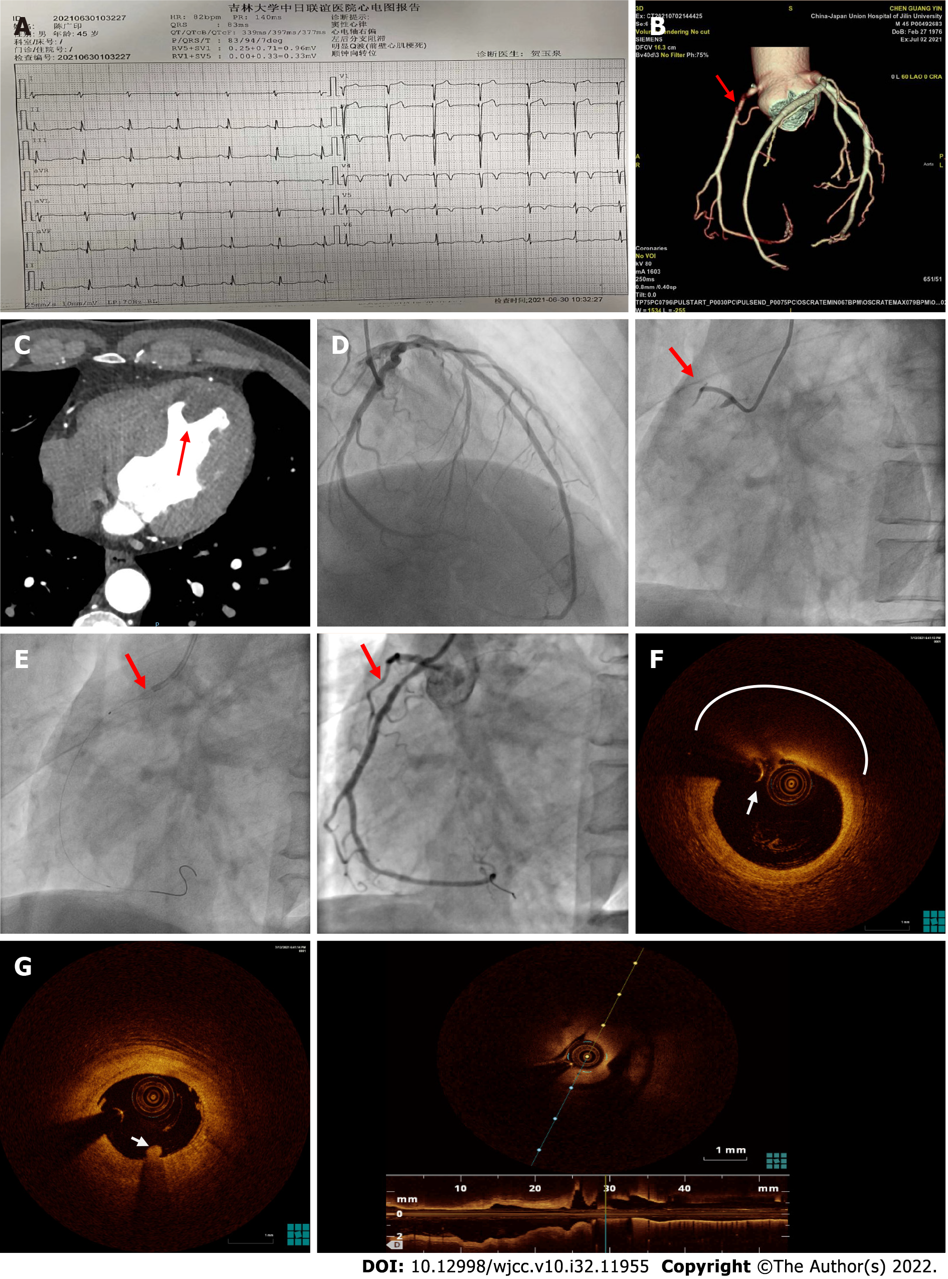
Figure 2 Second Admission electrocardiogram, coronary computed tomographic angiography, coronary angiography, and optical coherence tomography findings in 2021.
A: Admission electrocardiogram showed left posterior branch block and Q wave formation in anterior wall (red arrow); B: Coronary artery computed tomographic angiography (CTA) showed that the proximal segment of the right coronary artery was occluded by thrombosis, leading to moderate-to-severe stenosis in the corresponding lumen; C: Left ventricular thrombosis could be found in the CTA; D: Coronary angiography showed proximal segment of the right coronary artery occlusion; E: Coronary blood flow returned to TIMI 3 after embolization and aspiration; F: Optical coherence tomography results showed strong attenuation area with blurred edge and high dorsal reflection (white circle) could be seen at the proximal segment of the right coronary artery. The surface of the area was covered with a fibrous cap with high signal band (white arrow); G: Thrombus mass attached to the surface of the lumen could be observed (white arrow).
- Citation: Zhao YN, Chen WW, Yan XY, Liu K, Liu GH, Yang P. What is responsible for acute myocardial infarction in combination with aplastic anemia? A case report and literature review. World J Clin Cases 2022; 10(32): 11955-11966
- URL: https://www.wjgnet.com/2307-8960/full/v10/i32/11955.htm
- DOI: https://dx.doi.org/10.12998/wjcc.v10.i32.11955


