Copyright
©The Author(s) 2022.
World J Clin Cases. Nov 16, 2022; 10(32): 11955-11966
Published online Nov 16, 2022. doi: 10.12998/wjcc.v10.i32.11955
Published online Nov 16, 2022. doi: 10.12998/wjcc.v10.i32.11955
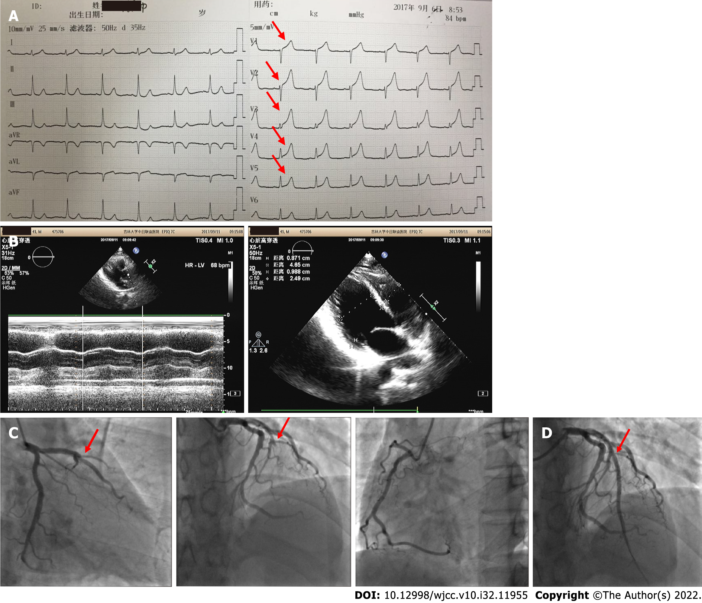
Figure 1 First admission electrocardiogram, coronary angiography, and echocardiography findings in 2017.
A: Admission electrocardiogram showed ST elevation and sharp T-wave in the anterior wall leads (red arrow); B: The motion amplitude of left ventricular anterior wall and anterior septum was weakened, and the ejection fraction was 46%; C: Emergency coronary angiography showed acute occlusion of the left anterior descending artery (red arrow); D: One stent was implanted for revascularization (red arrow).
- Citation: Zhao YN, Chen WW, Yan XY, Liu K, Liu GH, Yang P. What is responsible for acute myocardial infarction in combination with aplastic anemia? A case report and literature review. World J Clin Cases 2022; 10(32): 11955-11966
- URL: https://www.wjgnet.com/2307-8960/full/v10/i32/11955.htm
- DOI: https://dx.doi.org/10.12998/wjcc.v10.i32.11955


