Copyright
©The Author(s) 2022.
World J Clin Cases. Nov 16, 2022; 10(32): 11882-11888
Published online Nov 16, 2022. doi: 10.12998/wjcc.v10.i32.11882
Published online Nov 16, 2022. doi: 10.12998/wjcc.v10.i32.11882
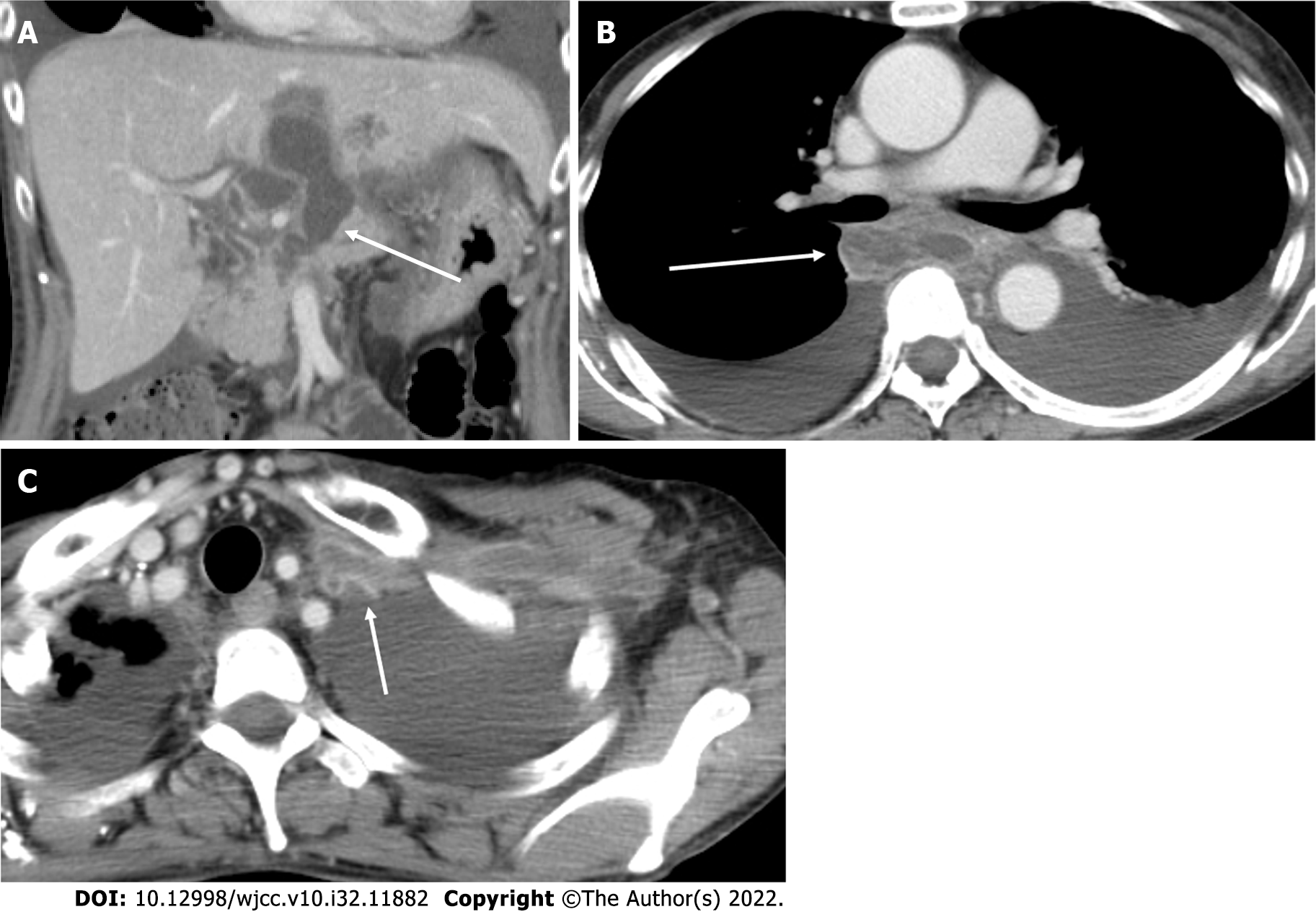
Figure 2 Contrast-enhanced computed tomography image.
A: Contrast-enhanced computed tomography image at the level of fluid collection around the head of the pancreas (arrow); B: Contrast-enhanced computed tomography image at the level of the mediastinal pseudopancreatic cyst (arrow); C: Contrast-enhanced computed tomography image at the level of brachiocephalic to brachial vein thrombotic vasculitis with contrast-enhancing vessel walls, suggesting inflammatory changes (arrow).
- Citation: Kokubo R, Yunaiyama D, Tajima Y, Kugai N, Okubo M, Saito K, Tsuchiya T, Itoi T. Brachiocephalic to left brachial vein thrombotic vasculitis accompanying mediastinal pancreatic fistula: A case report. World J Clin Cases 2022; 10(32): 11882-11888
- URL: https://www.wjgnet.com/2307-8960/full/v10/i32/11882.htm
- DOI: https://dx.doi.org/10.12998/wjcc.v10.i32.11882


