Copyright
©The Author(s) 2022.
World J Clin Cases. Nov 6, 2022; 10(31): 11542-11548
Published online Nov 6, 2022. doi: 10.12998/wjcc.v10.i31.11542
Published online Nov 6, 2022. doi: 10.12998/wjcc.v10.i31.11542
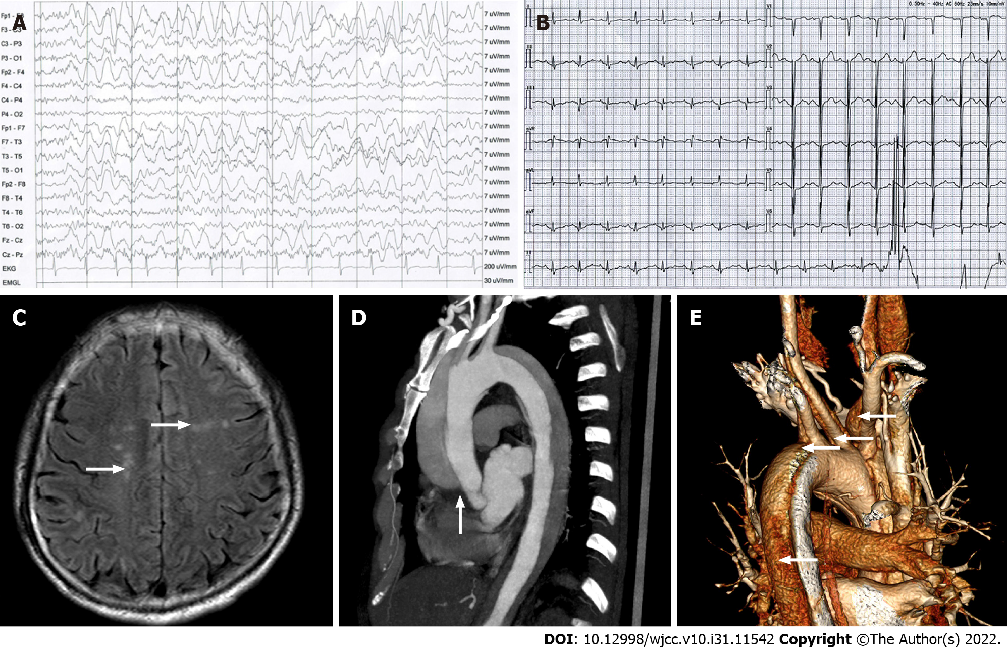
Figure 1 Examination results.
A: Electroencephalogram showed a lot of 2-3 Hz moderate-high amplitude slow waves; B: Electrocardiogram displayed sinus rhythm and there was no obvious abnormality; C: Head magnetic resonance imaging with FLAIR sequence (axial). Multiple abnormal hyperintense signals were seen in the centrum semiovale (white arrows), but there was no new infarction’s image characteristics around the precentral gyrus and other areas; D: Thoracic aorta computed tomography angiography [computed tomography angiography (CTA), left posterior]. The aortic dissection (AoD) originated at the lower part of the ascending aorta, with the true/false lumens on the right/Left sides of the white arrow, respectively; E: Three-dimensional reconstruction of the thoracic aorta CTA. The white arrows from the top-down suggested that the AoD involved the right common carotid artery, the brachiocephalic artery, the left common carotid artery, the left subclavian artery and the descending aorta.
- Citation: Zheng B, Huang XQ, Chen Z, Wang J, Gu GF, Luo XJ. Aortic dissection with epileptic seizure: A case report. World J Clin Cases 2022; 10(31): 11542-11548
- URL: https://www.wjgnet.com/2307-8960/full/v10/i31/11542.htm
- DOI: https://dx.doi.org/10.12998/wjcc.v10.i31.11542


