Copyright
©The Author(s) 2022.
World J Clin Cases. Jan 21, 2022; 10(3): 1131-1139
Published online Jan 21, 2022. doi: 10.12998/wjcc.v10.i3.1131
Published online Jan 21, 2022. doi: 10.12998/wjcc.v10.i3.1131
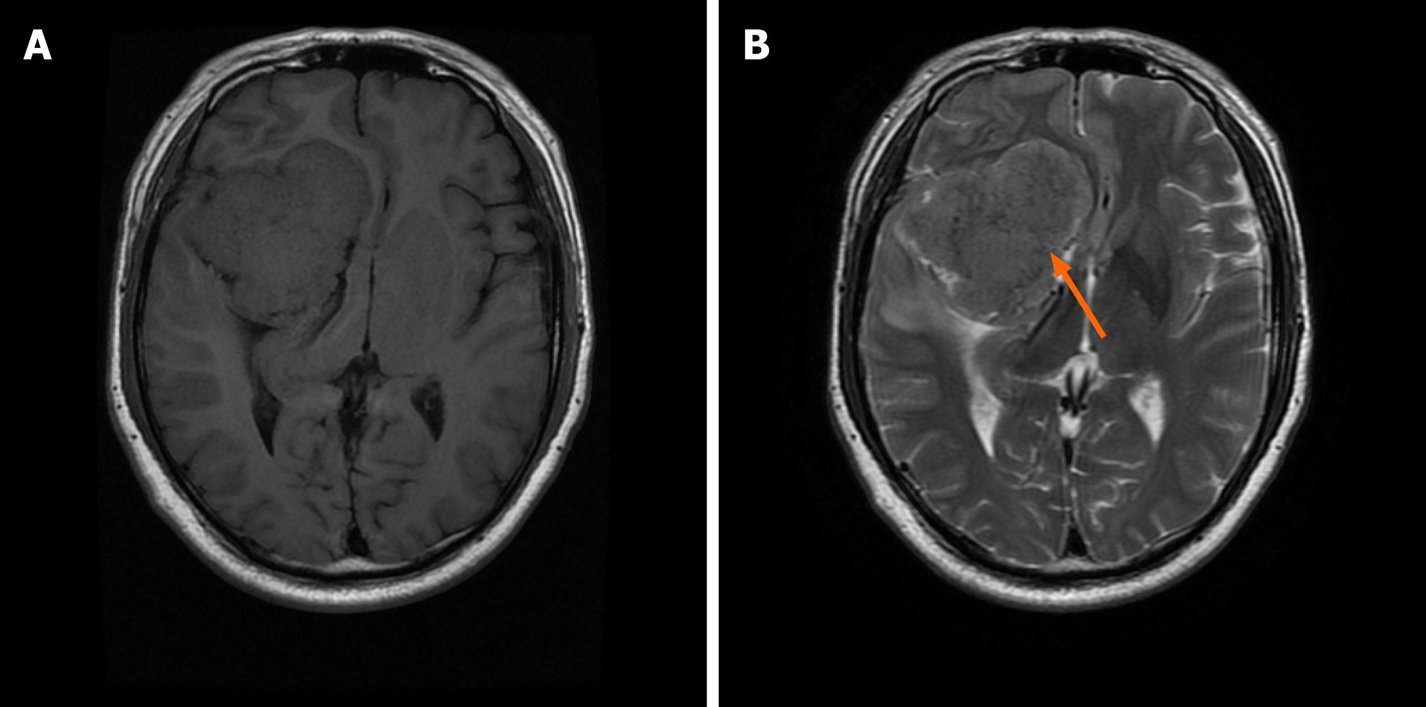
Figure 1 Magnetic resonance imaging scan of the brain.
T1- (A) and T2-weighted images (B) showed a large extracerebral mass at the right anterior, middle and posterior cranial base (orange arrow).
- Citation: Yang NL, Cai X, Que Q, Zhao H, Zhang KL, Lv S. Mycoplasma hominis meningitis after operative neurosurgery: A case report and review of literature. World J Clin Cases 2022; 10(3): 1131-1139
- URL: https://www.wjgnet.com/2307-8960/full/v10/i3/1131.htm
- DOI: https://dx.doi.org/10.12998/wjcc.v10.i3.1131


