Copyright
©The Author(s) 2022.
World J Clin Cases. Sep 6, 2022; 10(25): 8974-8979
Published online Sep 6, 2022. doi: 10.12998/wjcc.v10.i25.8974
Published online Sep 6, 2022. doi: 10.12998/wjcc.v10.i25.8974
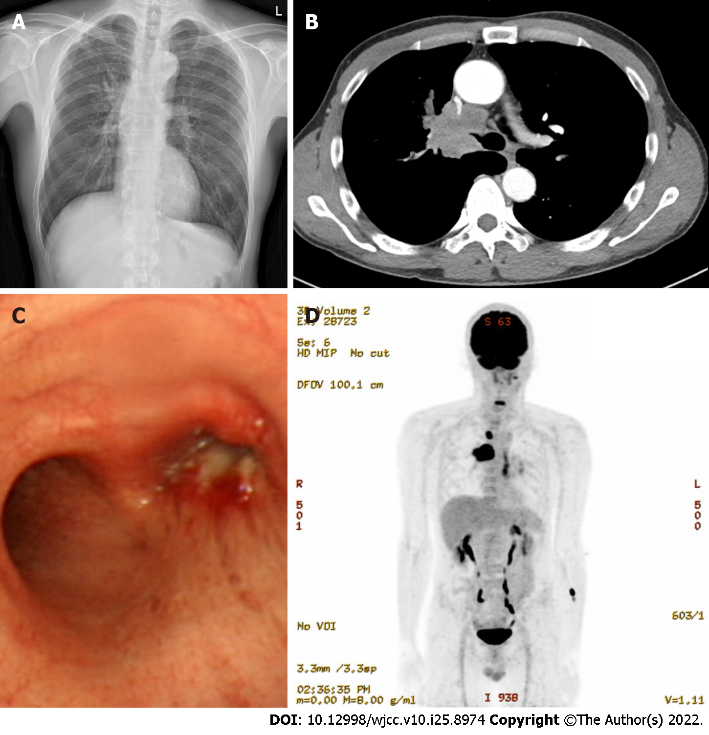
Figure 1 Initial chest radiography, chest computed tomography scan, bronchoscopy, and positron emission tomography-computed tomography.
A: Chest radiography reveals a mass-like shadow in the right supra-hilar area; B: Chest computed tomography (CT) showing narrowing of the right main bronchus secondary to a mass; C: Bronchoscopy confirms that the right bronchus is almost completely obstructed by a mass; D: Positron emission tomography-CT showing a 5-cm hypermetabolic mass (SUVmax 20.8) in the right upper lobe and hypermetabolic 4R and 2R lymph nodes.
- Citation: Yoo SS, Lee SY, Choi SH. Extracorporeal membrane oxygenation for lung cancer-related life-threatening hypoxia: A case report. World J Clin Cases 2022; 10(25): 8974-8979
- URL: https://www.wjgnet.com/2307-8960/full/v10/i25/8974.htm
- DOI: https://dx.doi.org/10.12998/wjcc.v10.i25.8974


