Copyright
©The Author(s) 2022.
World J Clin Cases. Aug 26, 2022; 10(24): 8474-8481
Published online Aug 26, 2022. doi: 10.12998/wjcc.v10.i24.8474
Published online Aug 26, 2022. doi: 10.12998/wjcc.v10.i24.8474
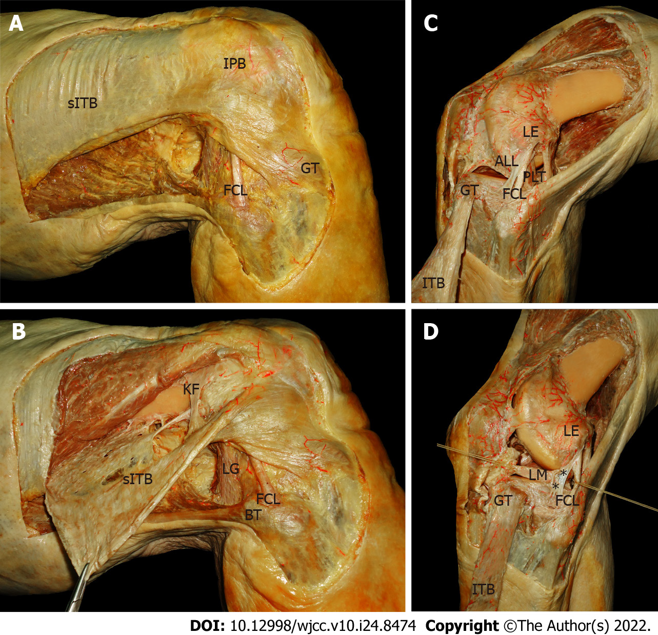
Figure 1 Photograph of anatomical dissection of right and left cadaver knees.
A and B: Right cadaver knee; C and D: Left cadaver knee. Asterisk: Coronary ligament which includes the meniscofemoral and meniscotibial ligament. sITB: Superficial iliotibial band; IPB: Iliopatelar band; GT: Gerdy’s tubercle; FCL: Fibular collateral ligament; KF: Kaplan fibers; BT: Biceps tendon; LG: Lateral gastrocnemius muscle; LE: Lateral epicondyle; ALL: Anterolateral ligament; ITB: Reflected iliotibial band; LM: Lateral meniscus.
- Citation: Garcia-Mansilla I, Zicaro JP, Martinez EF, Astoul J, Yacuzzi C, Costa-Paz M. Overview of the anterolateral complex of the knee. World J Clin Cases 2022; 10(24): 8474-8481
- URL: https://www.wjgnet.com/2307-8960/full/v10/i24/8474.htm
- DOI: https://dx.doi.org/10.12998/wjcc.v10.i24.8474


