Copyright
©The Author(s) 2022.
World J Clin Cases. Aug 16, 2022; 10(23): 8367-8374
Published online Aug 16, 2022. doi: 10.12998/wjcc.v10.i23.8367
Published online Aug 16, 2022. doi: 10.12998/wjcc.v10.i23.8367
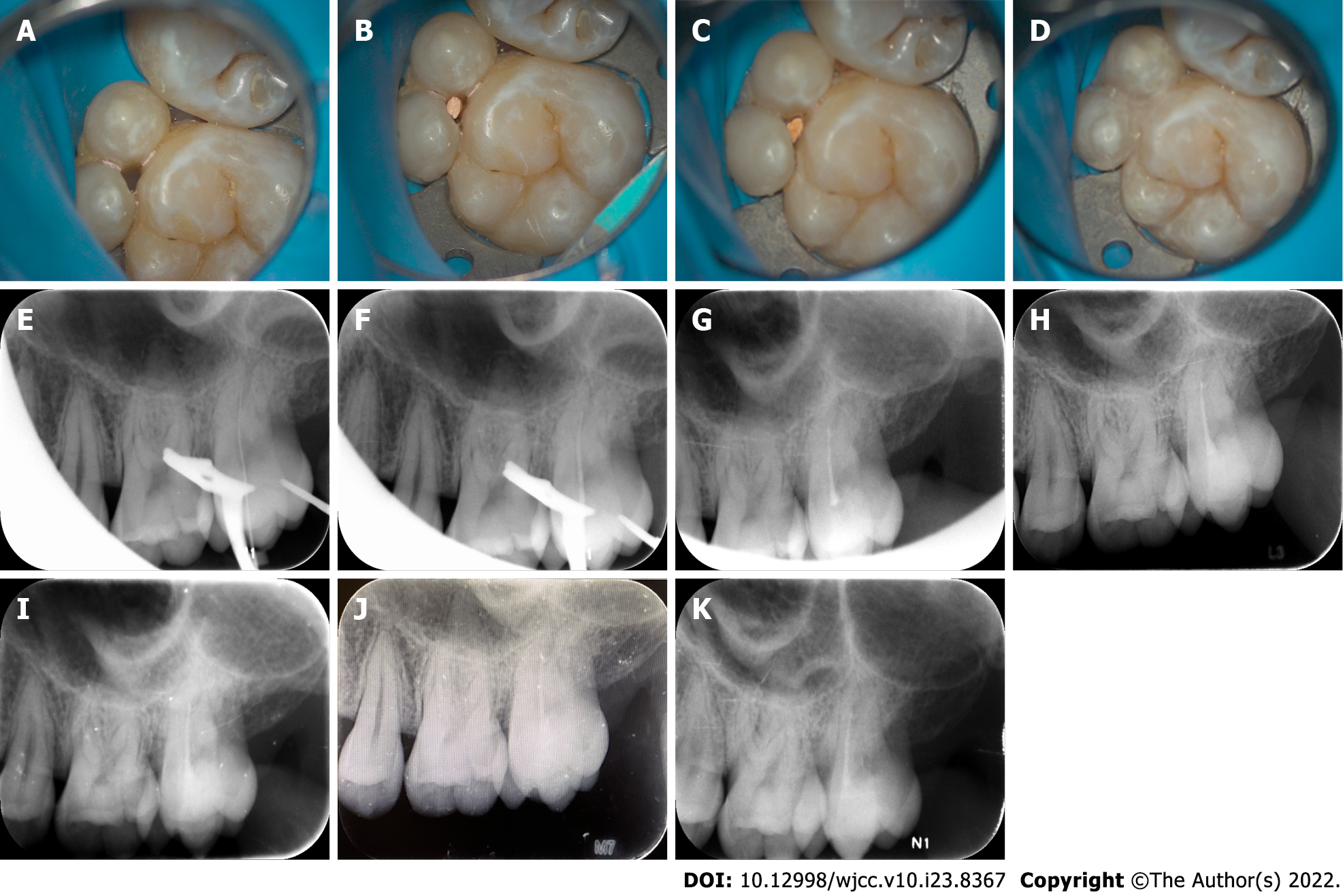
Figure 3 Representative intraoral photos of the fused tooth during root canal therapy and radiographs at different follow-up periods.
A: The canals were dried following a final irrigation; B: Occlusal view of the canal tip; C: Complete canal obturation was achieved via vertical gutta-percha condensation; D: Image of the nanoresin-mediated seal of the access cavity; E: Working length radiograph; F: Cone fit radiograph; G: A final digital PAX revealed that the canals were well-obturated; H-K: A radiograph taken during the 6-, 12-, 18-, 24-mo follow-ups revealed good treatment outcomes.
- Citation: Mei XH, Liu J, Wang W, Zhang QX, Hong T, Bai SZ, Cheng XG, Tian Y, Jiang WK. Endodontic management of a fused left maxillary second molar and two paramolars using cone beam computed tomography: A case report. World J Clin Cases 2022; 10(23): 8367-8374
- URL: https://www.wjgnet.com/2307-8960/full/v10/i23/8367.htm
- DOI: https://dx.doi.org/10.12998/wjcc.v10.i23.8367


