
















©The Author(s) 2022.
World J Clin Cases. Aug 16, 2022; 10(23): 8330-8335
Published online Aug 16, 2022. doi: 10.12998/wjcc.v10.i23.8330
Published online Aug 16, 2022. doi: 10.12998/wjcc.v10.i23.8330
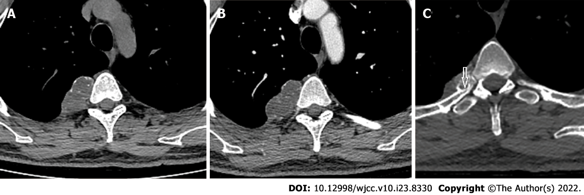
Figure 1 Computed tomography.
A and B: Representative plain computed tomography showed a lobulated soft tissue mass on the right side of the T4/5 vertebra that measured about 47 mm × 28 mm in the transverse view and contained diffuse stippled calcification; C: The mass caused cortical scalloping of the right fourth rib, rim ossification (arrow), and narrowing of the myeloid cavity. Enhanced computed tomography showed mild enhancement of the mass.
- Citation: Gao Y, Wang JG, Liu H, Gao CP. Periosteal chondroma of the rib: A case report. World J Clin Cases 2022; 10(23): 8330-8335
- URL: https://www.wjgnet.com/2307-8960/full/v10/i23/8330.htm
- DOI: https://dx.doi.org/10.12998/wjcc.v10.i23.8330