
















©The Author(s) 2022.
World J Clin Cases. Aug 6, 2022; 10(22): 8025-8033
Published online Aug 6, 2022. doi: 10.12998/wjcc.v10.i22.8025
Published online Aug 6, 2022. doi: 10.12998/wjcc.v10.i22.8025
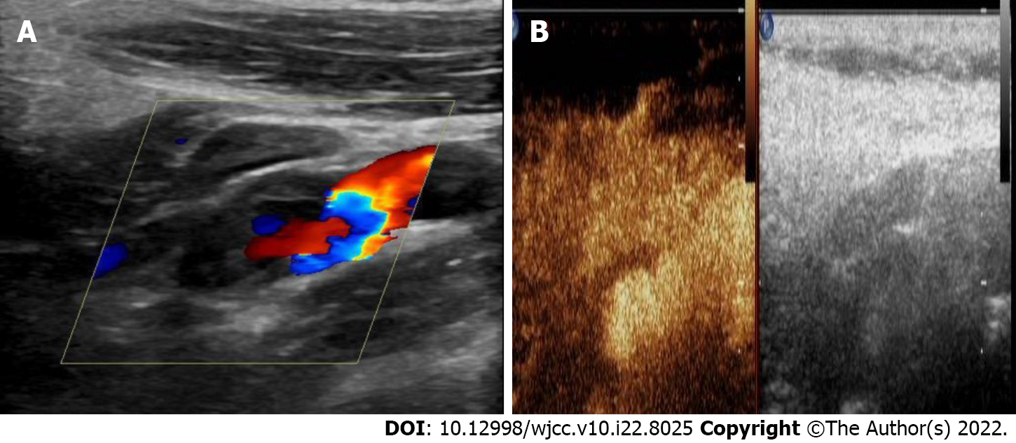
Figure 1 Ultrasonography and contrast-enhanced ultrasound of the carotid artery.
A: Conventional carotid artery ultrasonography showing a connection of the mass with the left internal carotid artery (ICA); B: Contrast-enhanced ultrasound of the carotid artery showing contrast agent filling in the distended area of the left ICA, but no enhancement in the low echo area of the mural.
- Citation: Zhong YL, Feng JP, Luo H, Gong XH, Wei ZH. Spontaneous internal carotid artery pseudoaneurysm complicated with ischemic stroke in a young man: A case report and review of literature. World J Clin Cases 2022; 10(22): 8025-8033
- URL: https://www.wjgnet.com/2307-8960/full/v10/i22/8025.htm
- DOI: https://dx.doi.org/10.12998/wjcc.v10.i22.8025