















©The Author(s) 2022.
World J Clin Cases. Aug 6, 2022; 10(22): 8009-8017
Published online Aug 6, 2022. doi: 10.12998/wjcc.v10.i22.8009
Published online Aug 6, 2022. doi: 10.12998/wjcc.v10.i22.8009
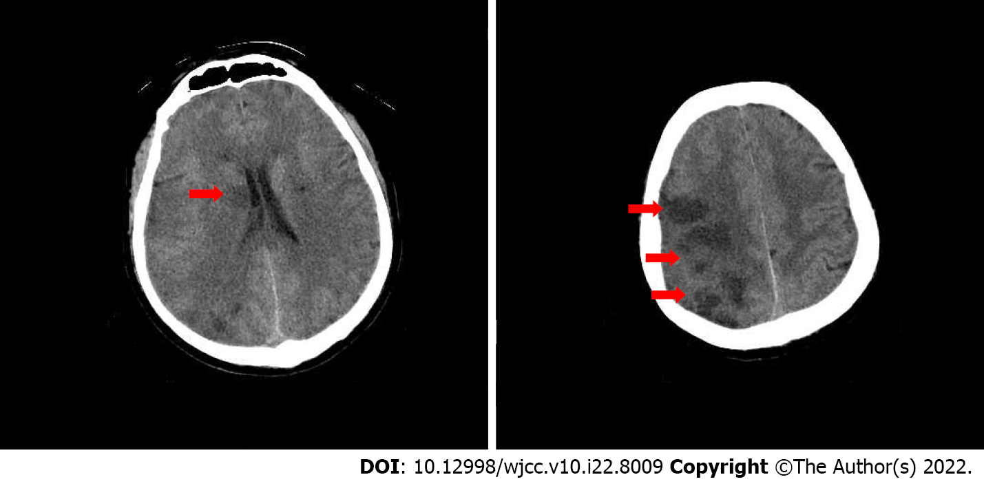
Figure 2 Cranial computed tomography on the second day of intravenous thrombolytic therapy.
Computed tomography shows right basal ganglia (A) and right frontotemporoparietal (B) brain tissue infarction (the red arrows).
- Citation: He ZY, Yao LP, Wang XK, Chen NY, Zhao JJ, Zhou Q, Yang XF. Acute ischemic Stroke combined with Stanford type A aortic dissection: A case report and literature review. World J Clin Cases 2022; 10(22): 8009-8017
- URL: https://www.wjgnet.com/2307-8960/full/v10/i22/8009.htm
- DOI: https://dx.doi.org/10.12998/wjcc.v10.i22.8009